Colour development protein chip using BCIP/NBT as substrate as well as application in detection of autoantibody thereof

A chromoprotein and chip technology, which is applied in biological testing, material inspection products, and analysis through chemical reactions of materials, to achieve high sensitivity and specificity, low detection cost, and easy promotion

Inactive Publication Date: 2008-07-23
SHANDONG MEDICAL BIO TECH RES CENT
View PDF0 Cites 13 Cited by
  • Summary
  • Abstract
  • Description
  • Claims
  • Application Information

AI Technical Summary

Problems solved by technology

However, the application of the NBT / BCIP colorimetric method in protein chips has not been reported at home and abroad so far. Therefore, it is necessary to combine this commonly used detection method with a new protein chip technology system to develop more suitable for different protein chips. A protein chip that is simple, fast, and simultaneously detects multiple antibodies or antigens required for level clinical testing

Method used


Image

  • Colour development protein chip using BCIP/NBT as substrate as well as application in detection of autoantibody thereof
  • Colour development protein chip using BCIP/NBT as substrate as well as application in detection of autoantibody thereof
  • Colour development protein chip using BCIP/NBT as substrate as well as application in detection of autoantibody thereof

Examples


Embodiment 1

[0029] Embodiment 1 takes BCIP / NBT as the chromogenic protein chip of substrate

[0030] 1. Preparation of APES-modified substrate ① Wash and dry the glass slide, soak in strong alkali (10M NaOH) for 24 hours; ② Rinse with deionized water 3 times, 10 minutes each time, then soak in strong acid (concentrated hydrochloric acid) 24 hours; ③ Rinse 3 times with deionized water, 10 minutes each time, spin dry in a centrifuge, stay in 2% APES acetone solution for 30-40 seconds, take it out and stop for a while, then rinse in pure acetone solution to remove untreated Combined APES, wash off excess acetone in deionized water; ④ Centrifuge to dry for later use, pay attention to dust.

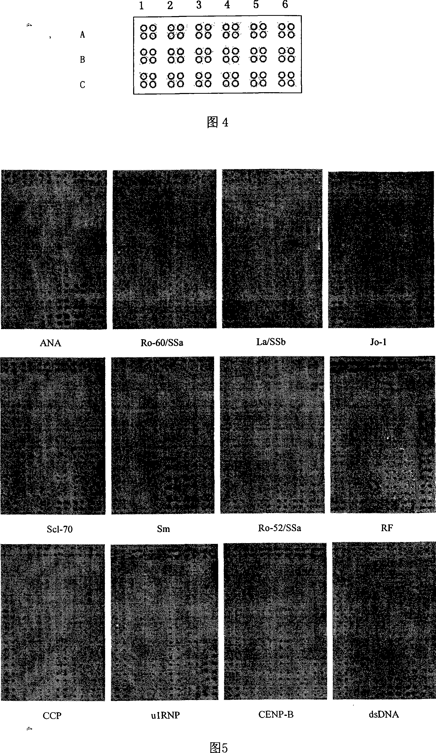
[0031] 2. Selection of antigen The present invention has selected 12 kinds of antigens commonly used in clinic for autoantibody detection, wherein Ro-52 / SSa, La / SSb, Jo-1 and Goat IgG are purchased from sigma; Ro-60 / SSa, Scl-70, CENP-B and u1RNP were purchased from Diarect; Sm was purchased from Immunovi...

Embodiment 2

[0038] Embodiment 2 optimization scheme selection test

[0039] 1. Optimization of spotting solution

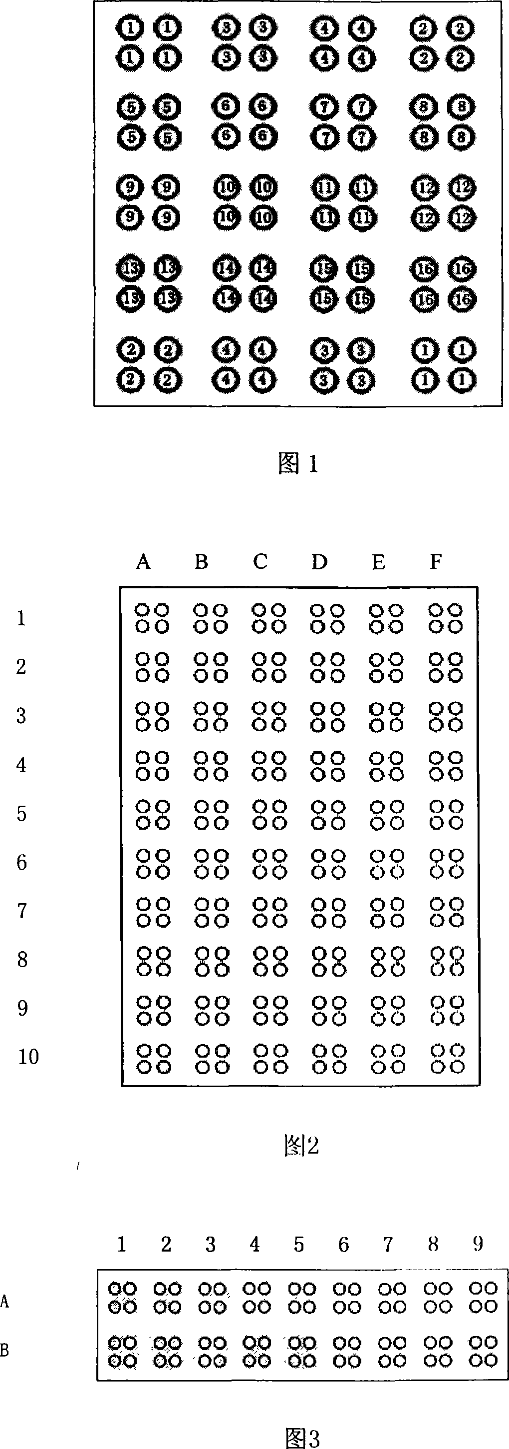
[0040] TBST containing different concentrations of Tween20 was selected as the spotting solution, the APES modified glass slide was used as the substrate, and samples were spotted with a Cartisian5500 spotting instrument. The array layout is shown in Figure 2. Each sample point was repeated 4 times, and the sample solution that could not only immobilize the antigen well but also improve the detection sensitivity was selected as the sample solution for the chip.

[0041] 2. Optimization of Serum Dilution

[0042] Dilute the standard antibody serum with PBS, the dilutions are 1:1, 1:2, 1:4, 1:8, 1:16, 1:32, on the same substrate, each array and a dilution Serum reaction; select the serum dilution with the highest signal-to-noise ratio as the serum reaction dilution of the chip.

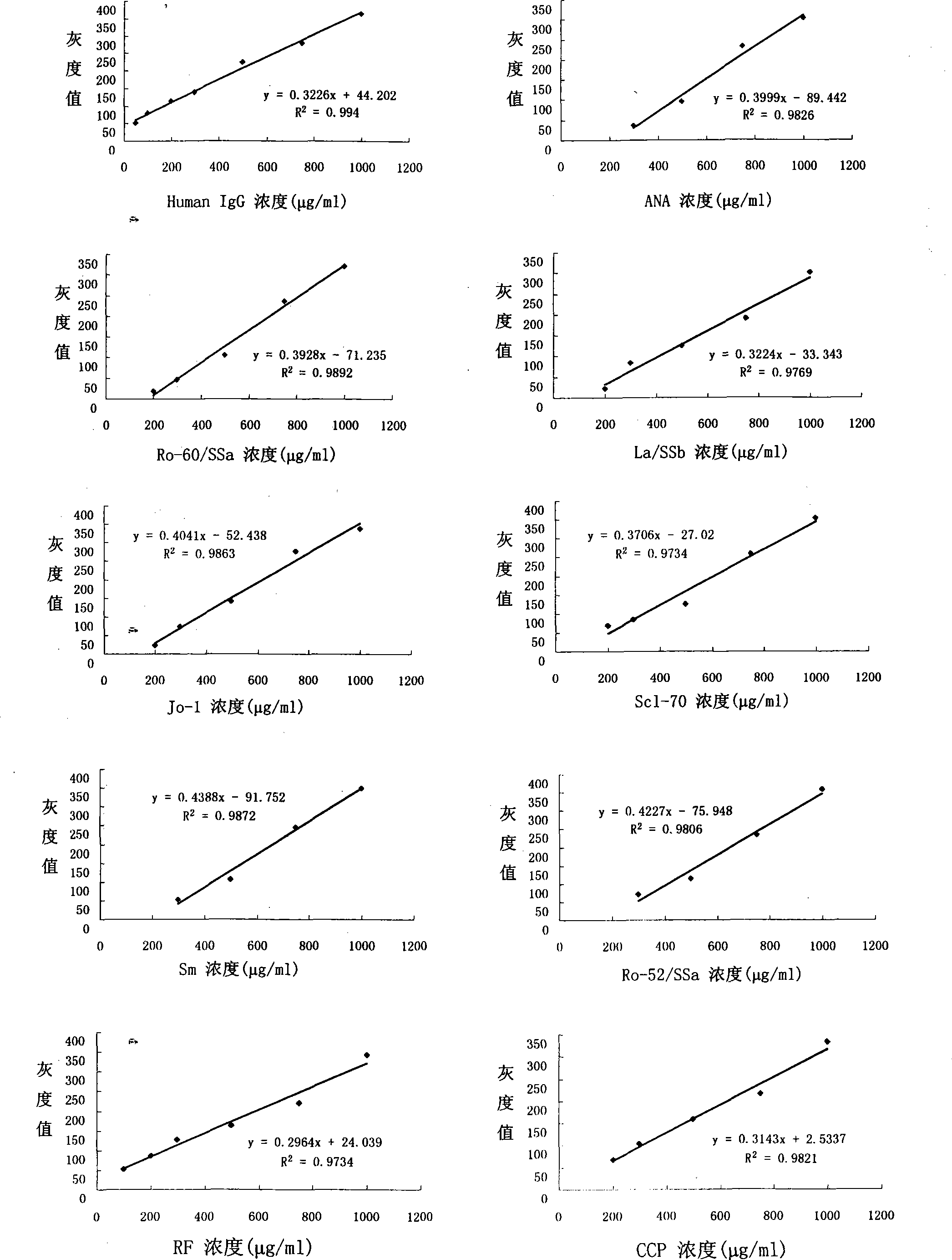
[0043] 3. Determination of spot concentration and positive cutoff value and negative cutoff valu...


PUM


Abstract

The invention discloses a color protein chip which substrate is BCIP/NBT. The invention is composed of a substrate, an array, 12 antigens coated on the array, negative contrast, positive contrast and blank contrast, wherein the substrate is a glass slide decorated by APES, the kinds and point sample densities of the 12 antigens are 520ug/ml ANA, 465ug/ml Ro-60, 530ug/ml La/SSb, 530ug/ml Jo-1, 525ug/ml Scl-70, 520ug/ml Sm, 615ug/ml Ro-52/SSa, 340ug/ml RF, 465ug/ml CCP, 410ug/ml u1RNP, 490ug/ml CENP-B, 580ug/ml dsDNA. The invention uses the alkali phosphatase coloring method whose substrate is BCIP/NBT as the final check signal of protein chip to be widely used for self antibody check, while the check sensitivity has higher coincidence rate than prior art, with reduced check cost and time. The invention can be used in clinical check, sanitary supervision, regular sanitary supervision and scientific research or the like.

Description

(1) Technical field [0001] The invention relates to a protein chip and its application, especially an alkaline phosphatase chromogenic protein chip with BCIP / NBT as a substrate and its application in autoantibody detection; it belongs to technologies such as biochip and diagnostic reagents field. (2) Background technology [0002] Autoimmune diseases (AID) can involve multiple organs and systems, causing great harm to patients. In recent years, the incidence of AID has increased significantly, accounting for about 3% to 5% of the world's total population, and there are more than 120 million patients in my country. The appearance of autoantibodies is the most important feature of AID. The detection of autoantibodies not only plays an important role in the early diagnosis of AID, but also has important significance in many aspects such as disease typing, prognosis judgment and treatment effect monitoring. [0003] At present, the clinical autoantibody detection methods mainl...

Claims


Application Information

Patent Timeline
Patent Type & Authority Applications(China)
IPC IPC(8): G01N33/53G01N21/78
Inventor 高雪芹王国强韩金祥
Owner SHANDONG MEDICAL BIO TECH RES CENT
Why Eureka
Try Eureka