Water relaxation-based sensors

A sensor and relaxation time technology, used in instruments, scientific instruments, magnetic variable measurement, etc., can solve the problem of nanoparticles not responding to the weak magnetic field of hand-held magnets

Inactive Publication Date: 2008-08-27
THE GENERAL HOSPITAL CORP
View PDF3 Cites 6 Cited by
  • Summary
  • Abstract
  • Description
  • Claims
  • Application Information

AI Technical Summary

Problems solved by technology

[0008] However, the nanoparticles do not r...

Method used


Image

  • Water relaxation-based sensors
  • Water relaxation-based sensors
  • Water relaxation-based sensors

Examples


Embodiment 1

[0191] Embodiment 1 Glu-CLIO nano-conversion

[0192] overview

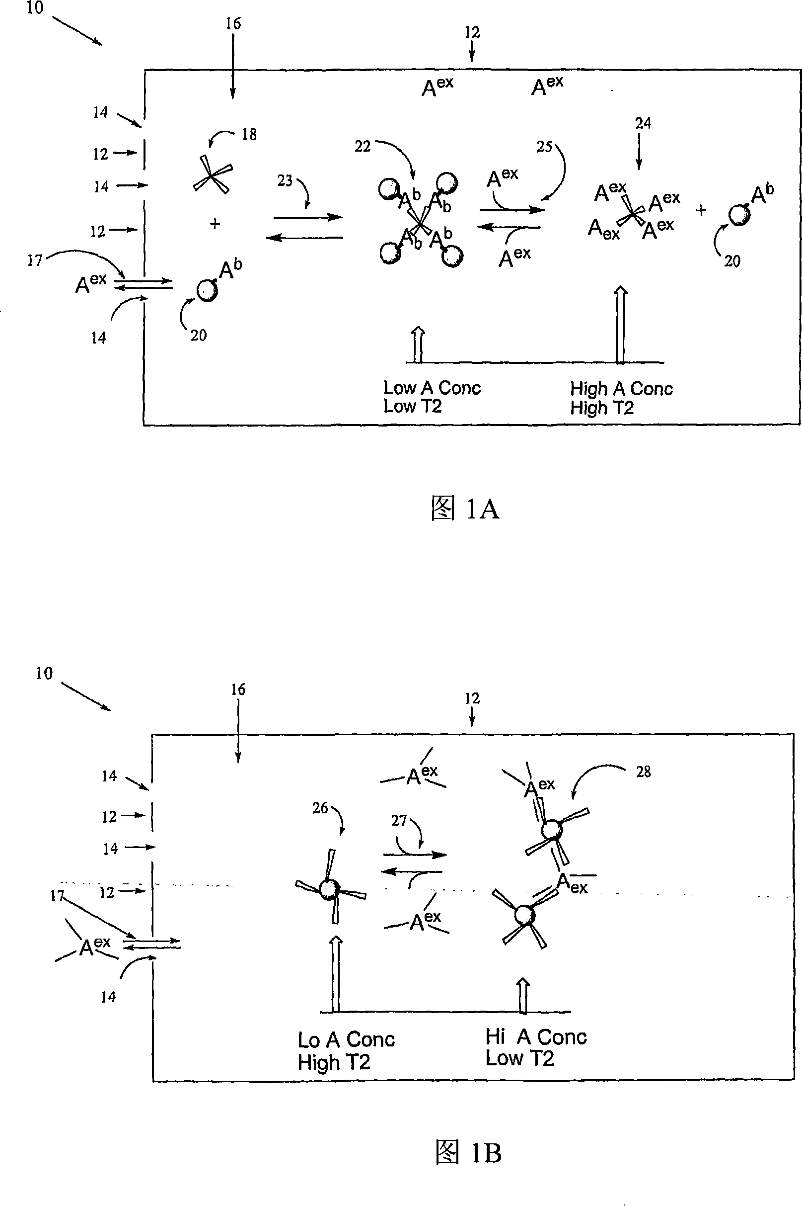
[0193] To demonstrate the water relaxation sensor, we designed a model machine for monitoring physiological concentrations of glucose. We used (con)conavalin A as binding protein and synthetic glucose-functionalized magnetic nanoparticles (Glu-CLIO). Concanavalin A (ConA) is a tetravalent lectin known to react with glucose. Some sensors were prepared with wall shells with a pore size of 3 kDa. In general, the wall shell of the sensor holds the Glu-CLIO nanoparticles and ConA, while allowing glucose to freely pass in and out of the sensor.

Embodiment 1A

[0194] Preparation of Example 1A.Glu-CLIO

[0195] MION-47 and amino-CLIO (25-35 nm) were prepared as described elsewhere. D-glucose, D-(+)-glucosamine hydrochloride, succinic anhydride, (con)concanavalin A (ConA) and Sephadex G-25 were from Sigma Aldrich. 1-Ethyl-3-(3-dimethylaminopropyl)carbodiimide hydrochloride (EDC) and sulfo-N-hydroxysuccinimide (sulfo-NHS) were from Pierce (Rockford, IL). In order to synthesize glucose-functionalized nanoparticles (Glu-CLIO), the NH 2 -CLIO was transformed into carboxyl-functionalized nanoparticles, which were then coupled to 2-amino-glucose using water-soluble carbodiimide. To obtain carboxy-functionalized CLIO, add 2.0 mg succinic anhydride to 200 uL NH 2 -CLIO (10mgFe / mL, 42NH 2per 2064 Fe) with 300uL (0.1M) NaHCO 3 buffer, pH 8.5. The mixture was incubated for two hours at room temperature and eluted with MES buffer (0.5M NaCl, 0.05MMES), pH 6.0, using a Sephadex G-25 column to remove succinic acid. To conjugate 2-amino-gluc...

Embodiment 1B

[0196] Example 1B. Analysis of Glu-CLIO-ConA-Glucose Tubes

[0197] Relaxation times were obtained using a Minispec stress relaxometer (Bruker) at 0.47T, 40°C.

[0198] To demonstrate the interaction of Glu-CLIO, ConA and glucose, experiments were performed directly in NMR tubes (without semipermeable wall enclosures), 10ugFe / mL, 800ug / mL ConA. All experiments were performed with lmM CaCl 2 and 1mM MgCl 2 performed in PBS. Transverse relaxation times (T2's) were measured with a stress relaxometer Bruker Minispec(R) NMS 120 at 0.47T and 40°C. Size was determined with a Zetasizer 1000(R) (Malvern Instruments, Marlboro, MA) in the above buffer with Glu-CLIO at 20 ug Fe / mL, followed by addition of ConA to 1 mg / mL and 1.5 mg / mL glucose. These concentrations of ions, buffer, ConA and Glu-CLIO were used in all experiments.

[0199] As shown in Figure 3A, addition of ConA to Glu-CLIO decreased T2, which reached a plateau after about 50 min, while there was no change for amino-CLI...


PUM


Abstract

The present invention relates to magnetic resonance-based sensors and related methods . More specifically, the invention relates to a sensor based on magnetic resonance for detecting the presence of an analyte in a sample solution. The sensor comprises magnetic nanoparticles linked to a moiety upon which the analyte can reversibly bind, the nanoparticles being confined in a chamber with semipermeable walls which are impermeable to the nanoparticles but permeable to the analyte. Upon binding of the analyte to the nanoparticles, the latter aggregate which can be deteceted by a change in the T2 relaxation time of the sample solution within the chamber (magnetic relaxation switch) . The analyte may be a carbohydrate such as glucose, an antibody, an amino acid, a nucleic acid, an oligonucleotide, a therapeutic agent, a peptide, a protein, etc. The sensor may be used as an assay and may be read out using MRS or MRI, e.g. of well plates for high throughput anaylsis .

Description

[0001] Cross reference to related applications [0002] This application claims priority to US Provisional Application 60 / 679,437, filed May 9, 2005, which is hereby incorporated by reference in its entirety. technical field [0003] The present invention relates to magnetic resonance based sensors and related methods. [0004] Notes on Research Initiated on a Commonwealth-wide basis [0005] The work described here was performed at least in part using grants RO 1EB 004626 and EB 00662 from the National Institutes of Health (NIH) grants. The Government therefore has certain rights in this invention. Background technique [0006] Magnetic resonance (MR) based recording methods, such as magnetic resonance imaging (MRI), have certain known advantages as non-invasive methods. For example, magnetic resonance imaging can be used deep in tissue where optical recording methods are sometimes complicated by light scattering and absorption by the tissue, eg, deeper than about 250 mi...

Claims


Application Information

Patent Timeline
IPC IPC(8): G01R33/465
CPCA61B5/14503G01R33/465G01N33/54366G01N33/54326G01R33/50G01N27/745
Inventor 李·约瑟夫森孙易拉尔夫·魏斯勒德
Owner THE GENERAL HOSPITAL CORP
Why Eureka
Try Eureka