
Paragonimiasis detection device and method for making same

A detection device and a technology for paragonimiasis, which are applied in measurement devices, biological tests, material inspection products, etc., can solve problems such as cross-reaction interference, unsatisfactory effect of curative effect assessment, comparison of test results, etc., and achieve less cross-reaction interference and detection Fast, low-cost detection results

Inactive Publication Date: 2008-09-17
ARMY MEDICAL UNIV
View PDF1 Cites 6 Cited by
  • Summary
  • Abstract
  • Description
  • Claims
  • Application Information

AI Technical Summary

Problems solved by technology

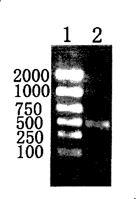
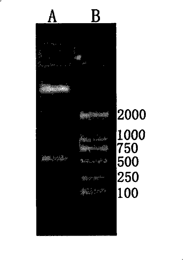
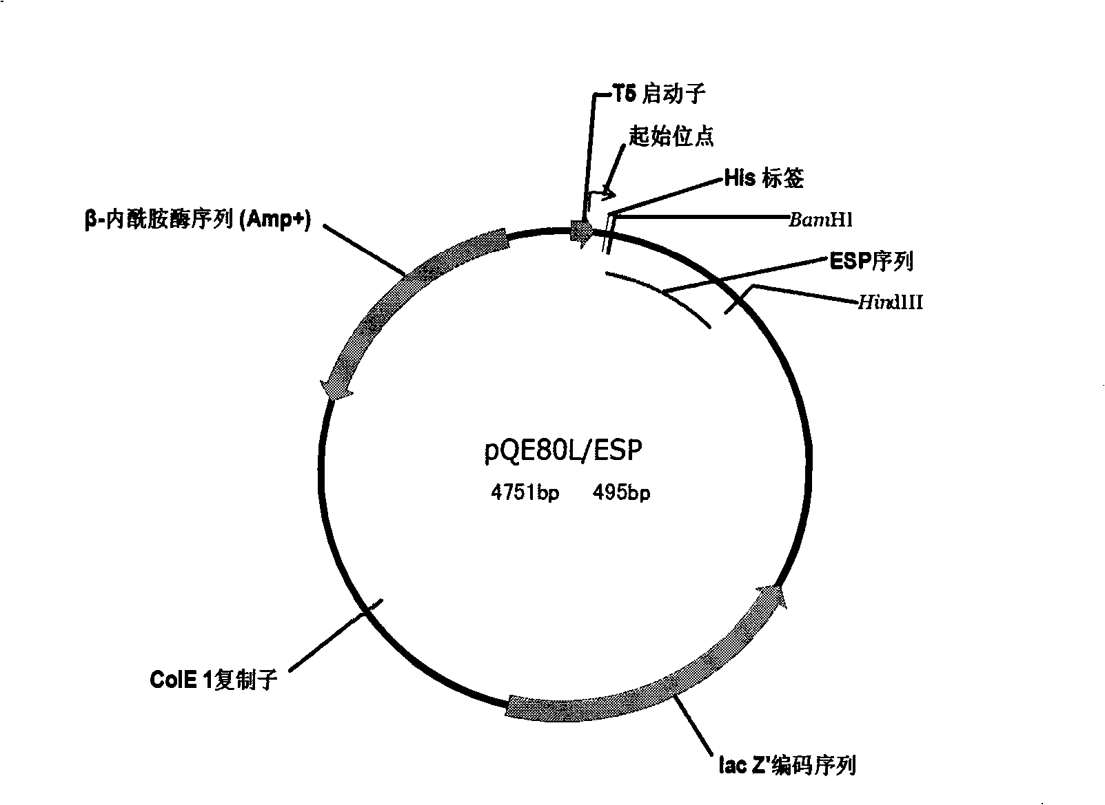
[0005] In view of this, in order to solve the technical problems such as unsatisfactory curative effect assessment, cross-reaction interference, and inability to compare the test results of various laboratories in the antibody detection method of the existing paragonimiasis immunological detection method, the inventor adopted Sodium dodecyl sulfate-polyacrylamide gel electrophoresis (SDS-PAGE), two-dimensional gel electrophoresis (2-DE), western blotting, mass spectrometry Analyzing the parasite protein at the stage, identifying the specific antigen that can be used for immune detection in the parasite excretion, and obtaining its coding gene fragment by reverse transcription polymerase chain reaction (RT-PCR) (Genbank accession number is AY083923 ), the polypeptide deduced according to the obtained sequence is composed of 165 amino acids, named as Excretory-secretory protein (Excretory-secretory protein, ESP), the protein is prepared and the specific antibody is prepared through animal immunization, and then the paragonimiasis is further obtained Detection device

Method used


Image

  • Paragonimiasis detection device and method for making same
  • Paragonimiasis detection device and method for making same
  • Paragonimiasis detection device and method for making same

Examples


Embodiment Construction

[0035] The present invention will be described in detail below with reference to preferred embodiments. It should be understood that the following preferred embodiments are only for illustrating the present invention, rather than limiting the protection scope of the present invention.

[0036] 1. Preparation of Paragonimiasis Detection Device

[0037] 1. Preparation of ESP protein from Paragonimus sinensis

[0038] a. Design and synthesis of primers

[0039] According to the conserved amino acid sequence of trematode cysteine ​​protease, the following degenerate primers were designed and synthesized:

[0040] Upstream primer: 5'-TCA(AG)GG(AGCT)CA(AG)TG(CT)GG(AGCT)TC(AGCT)TG(CT)TGG-3';

[0041] Downstream primer: 5'-CCA(AG)CT(AG)TT(CT)TT(AGCT)AC(AG)ATCCA(AG)TA-3';

[0042] b. RT-PCR amplification of the ESP protein coding gene of Lung fluke

[0043] Get 2 adult worms (60 mg in total) of U. skrenzii, homogenize them in an ice bath with Tripure reagent (Roche, USA), and extr...


PUM


Abstract

The invention discloses a device for detecting paragonimiasis and preparation method, comprising a label pad, a detecting layer, wherein the labelled detection antigen is on the labelled pad and the detection antigen is the lung fluke ESP protein represented by the amino acid sequence SEQ ID NO.2. The preparation method comprises preparing the lung fluke ESP protein and polyclonal antibody thereof, and preparing the label pad, the detection layer, the sample pad, the absorption pad and the detection device. The detection device prepared by the preparation method has features: high specificity, sensitivity, small cross reaction with the clonorchiasis and schistosomiasis, false positive rate lower than ELISA method, with curative effect evaluating function. The detection result becomes negative in 3-6 monthes after curing the paragonimiasis by effective medicine and the negative rate is higher than the ELISA method and the device for detecting paragonimiasis can accurately, quickly, simply perform diagnosis, differential diagnosis and evaluation of the curative effect with important clinical application value.

Description

technical field [0001] The invention relates to a detection device, in particular to a paragonimiasis detection device and a preparation method thereof. Background technique [0002] Paragonimiasis (Paragonimiasis) is a common and important zoonotic parasitic disease, mainly prevalent in Asia, Africa and Latin America, especially in Asia, prevalent in 22 provinces, municipalities and autonomous regions in my country, It is estimated that about 2 million people are infected every year, seriously endangering human health, especially the health of adolescents and children. [0003] Paragonimiasis is mainly caused by mechanical damage caused by paragonimiasis (larvae or adults) migrating and living in human tissues and organs, and immunopathological reactions caused by their metabolites. In my country, the pathogenic parasites are mainly lung fluke wescheri and lung fluke steineri, and their adults parasitize in the muscles and organs of carnivorous animals such as humans, dogs,...

Claims


Application Information

Patent Timeline
IPC IPC(8): G01N33/68G01N33/558G01N33/532
Inventor 张锡林
Owner ARMY MEDICAL UNIV
Learn More