
Method and device of dual-color single photon transverse super resolution imaging

A technology of super-resolution imaging and imaging device, which is applied in measurement devices, material analysis by optical means, and material analysis, etc., can solve problems such as expensive equipment and complicated processes, and achieve the effect of convenient operation and high fluorescence excitation efficiency.

Inactive Publication Date: 2009-05-20
SHANGHAI INST OF OPTICS & FINE MECHANICS CHINESE ACAD OF SCI
View PDF0 Cites 18 Cited by
  • Summary
  • Abstract
  • Description
  • Claims
  • Application Information

AI Technical Summary

Problems solved by technology

The principle of energy transfer resonance between fluorescent molecules and single fluorescent molecular positioning technology can also break through the diffraction limit, but these technologies often require very harsh conditions and expensive equipment, and the process is also very complicated

Method used


Image

  • Method and device of dual-color single photon transverse super resolution imaging
  • Method and device of dual-color single photon transverse super resolution imaging
  • Method and device of dual-color single photon transverse super resolution imaging

Examples


Embodiment 2

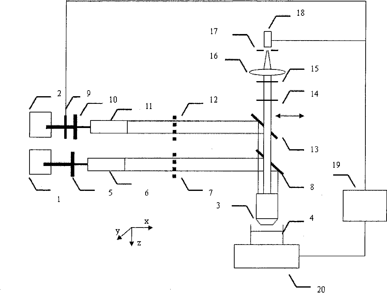
[0049] Optical path diagram such as figure 1 As shown, the sensitizing pupil 7 and the excitation pupil 12 are not added in the optical path, and the excitation dichroic mirror 13 is adjusted in the x-axis direction so that the two beams of light propagate in parallel and non-coaxially, but the two beams of light spot spread after being focused by the objective lens 3 The function center is at a distance of L=220nm in the x-axis. At this time, the fluorescence excitation of the reversible photosensitive fluorescent protein can only be realized in the overlapping region of the edge of the airy spot of the sensitizing light and the excitation light, and other non-overlapping regions cannot meet the fluorescence excitation conditions of the reversible photosensitive fluorescent protein without emitting fluorescence. The equivalent point spread function is as Figure 6 As shown, the x-axis full width at half maximum is 79nm, and the resolution in the x direction is increased by 2...

Embodiment 3

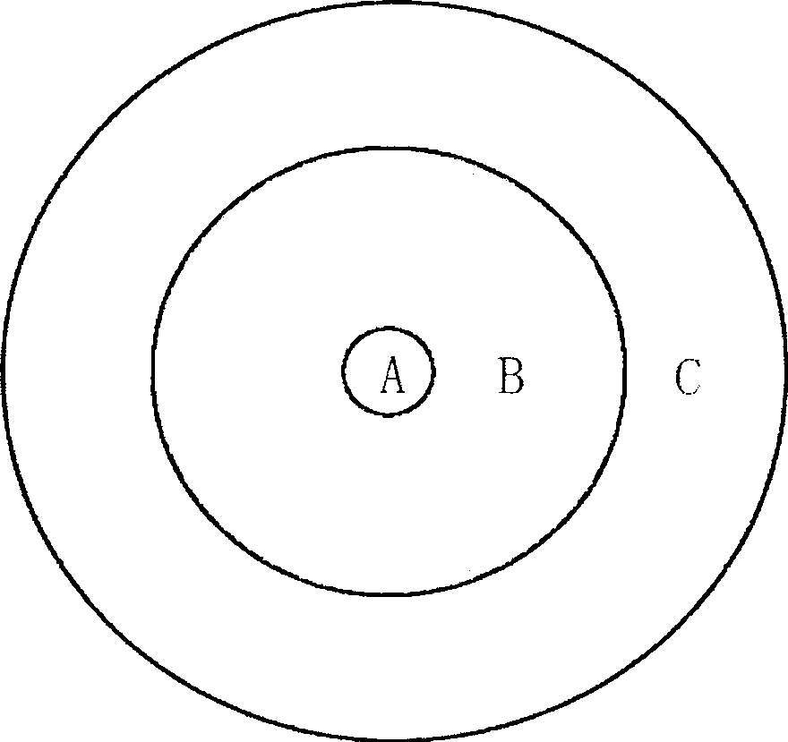
[0051] Optical path diagram such as figure 1 As shown, a sensitization pupil 7 and an excitation pupil 12 are added to the optical path, and the sensitization pupil 7 and the excitation pupil 12 are three-zone phase pupil filters, with three zones A, B, and C from inside to outside The phases are 0, π, 0 in sequence, and the normalized radius is 0.12:0.6:1 in sequence. The addition of the pupil changes the distribution of the original point spread function and enhances the maximum peak of the third-order diffraction. By adjusting the excitation dichroic mirror 13 in the x-axis direction, the two beams of light propagate in parallel and non-coaxially, and after being focused by the objective lens 3, the x-axis distance of the center of the point spread function of the two beams of light is L=1010 nm. At this time, the fluorescence excitation of the reversible photosensitive fluorescent protein can only be realized in the overlapping area of ​​the focus edge of the third-order ...


PUM

PropertyMeasurementUnit
Wavelengthaaaaaaaaaa

Abstract

The invention provides a dual-color single-photon transverse super-resolution imaging method, and a device thereof. The method has the most important characteristic that fluorescence excitation can be achieved only by adopting a overlapping part of a sensitization beam and an excitation beam to simultaneously sensitize and excite samples labeled with reversible photosensitive fluorescent protein molecules. The method comprises the following steps: fluorescence signals are collected by an objective, pass a sensitization dichroic mirror, an excitation dichroic mirror, a notch light filter and a long-channel light filter, and are focused by a focusing lens to pass a pinhole aperture; fluorescence intensity is measured by an avalanche diode; fluorescence intensity values are recorded by a computer; the computer closes a shutter so as to restore reversible photosensitive fluorescent protein to an excitable state under the illumination of sensitization light; and the computer drives a 3-D translation stage to move the samples labeled with the reversible photosensitive fluorescent protein, so as to achieve 3-D scanning imaging. The method can improve the transverse resolution of super-resolution fluorescence imaging by 1.55 to 2.81 times.

Description

technical field [0001] The invention relates to biomedicine, and relates to two-color single-photon fluorescence imaging, in particular to a method and device for two-color single-photon lateral super-resolution imaging, which is suitable for the detection of medical biological tissues. Background technique [0002] For a long time, far-field optical fluorescence microscopy has been the most commonly used observation tool in life sciences due to its advantages of non-contact, non-damage, and ability to detect the interior of samples. However, due to the existence of the diffraction limit, the lateral resolution of the traditional wide-field optical microscope is only about 200nm. In order to reveal the dynamic and structural characteristics of intracellular molecular scale, improving the resolution of optical microscopy has become an urgent requirement for the development of life sciences. On the basis of far-field fluorescence microscopy, scientists have developed some pra...

Claims


Application Information

Patent Timeline
IPC IPC(8): G01N21/64G01N33/48
CPCG01N21/6428G01N21/6458G01N2021/6419
Inventor 毛峥乐王琛乔玲玲程亚
Owner SHANGHAI INST OF OPTICS & FINE MECHANICS CHINESE ACAD OF SCI
Learn More