
X ray perspective view calibration method in operation navigation system

A technique of surgical navigation and calibration method, which is applied in the field of X-ray fluoroscopic image calibration in a spinal surgery navigation system, can solve the problems of limiting the functional application of a C-arm X-ray machine, affecting the flexibility of surgical navigation, and the stability to be improved, etc. To achieve the effect of convenient clinical application, difficult to estimate deformation, and reliable algorithm

Inactive Publication Date: 2009-07-01
FUDAN UNIV +1
View PDF0 Cites 36 Cited by
  • Summary
  • Abstract
  • Description
  • Claims
  • Application Information

AI Technical Summary

Problems solved by technology

However, this type of method has the following disadvantages: First, surgical navigation can only be carried out in the fluoroscopy position where the internal parameters of the imaging system are fixed, which limits the functional application of some C-arm X-ray machines and affects the flexibility of surgical navigation.
However, due to the interference of images such as the patient's body and surgical instruments in addition to the marker point information in the fluoroscopic image, the calibration process is complicated and the stability needs to be improved.

Method used


Image

  • X ray perspective view calibration method in operation navigation system
  • X ray perspective view calibration method in operation navigation system
  • X ray perspective view calibration method in operation navigation system

Examples


Embodiment 1

[0019] 1. X-ray fluoroscopy image acquisition and preprocessing

[0020] Use video acquisition technology to digitize X-ray perspective images, and use multi-frame average filtering and wavelet transform methods to comprehensively suppress noise. Firstly, multiple images are superimposed and averaged, and then the averaged image is used as the input image for wavelet transform filtering. The type of wavelet adopts the symmetric wavelet symN proposed by Daubechies, N=2,3,...8, and adopts soft thresholding and fixed threshold criterion for denoising. After the above series of processing, the image noise is effectively filtered out and the detailed information in the original image is better preserved.

[0021] 2. Mark point recognition and correction

[0022] First, filter the background information. Using the principle of digital subtraction: set the original image as I Source , Using the kernel function with the diameter of the marker point as the size to compare I Source Perform ...

Embodiment 2

[0085] Example 2 Collection and calibration test of clinical fluoroscopy images

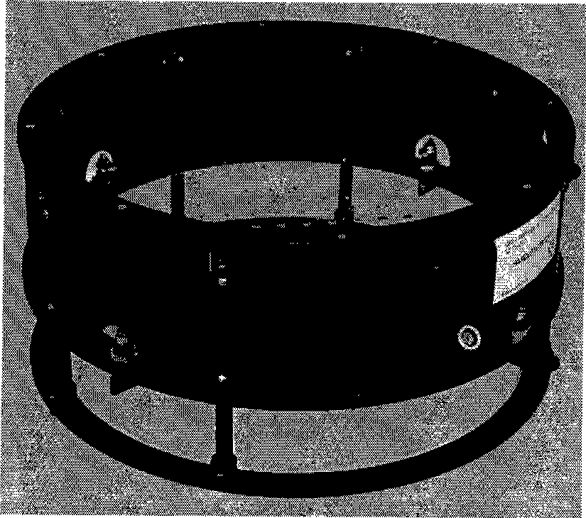
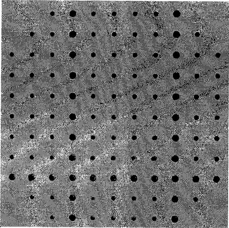
[0086] The calibration target is fixed on the image intensifier of the C-shaped wall X-ray machine, and the proximal target surface is close to the input screen of the intensifier to collect the patient's fluoroscopy image. Taking into account the sampling time and the movement of the patient's body, the present invention superimposes and averages the images collected within 0.5 seconds after the fluoroscopic pedal is released, and performs wavelet filtering;

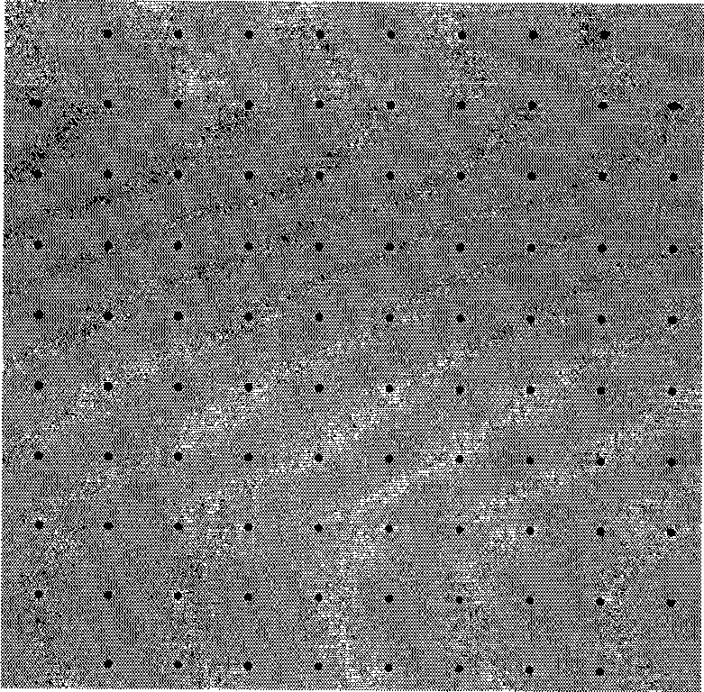
[0087] Automatically recognize the marked points, the kernel function of the median filter is 2.5mm; the present invention uses filtering and subtraction methods to remove the information of non-marked points in the image. In the template matching algorithm, with 0.85 as the threshold, all points with R(i,j)>0.85 are listed as possible marker point centers, clustering is performed to find the class center, and then the marker is obtained by th...


PUM


Abstract

The invention belonging to process and application field of medical images relates to an X-ray perspective image demarcating method of an operation navigation system. The method comprises obtaining X-ray perspective images containing marking point information in the operation, performing filtration and subtraction to obtain the images only containing marking point information, obtaining the center coordinate and the arrangement direction of the marking points by utilizing methods of template matching, cluster analysis and statistics of information entropy, correcting the marking point coordinate by using a B belt transect and calculating the demarcating parameters. A projection coordinate of the marking point is obtained according to the obtained imaging model parameters, all pixels of the image is corrected according to the distance between the projection coordinate and the real coordinate on the image to obtained a corrected X-ray perspective image for the operation navigation system. The method has characteristics of simple embodiment, reliable algorithm, convenience in clinical application and improvement of precision of the operation navigation system.

Description

Technical field [0001] The invention belongs to the field of medical image processing and application, and relates to a method for X-ray fluoroscopy image calibration, in particular to an X-ray fluoroscopy image calibration method in a spinal surgery navigation system. Background technique [0002] Surgical navigation based on C-arm X-ray fluoroscopy images is a main type of spine surgery navigation, which is mostly used for thoracolumbar spine surgery. For this type of surgical navigation, the internal parameters of the imaging system must be calculated based on the X-ray fluoroscopy image, such as focal length, image scale factor, radial distortion, etc., as well as external parameters, such as focus space position, projection direction, etc. The above process is called the calibration of the imaging system. At the same time, due to the spherical effect of the image intensifier of the X-ray machine and the magnetic field offset caused by gravity, the obtained perspective image ...

Claims


Application Information

Patent Timeline
IPC IPC(8): A61B6/02A61B19/00G06T7/00A61B34/20
Inventor 张翼宋志坚王满宁李文生姚德民
Owner FUDAN UNIV
Learn More