Method for constructing oral squamous cell carcinoma animal model
An animal model, oral mucosal technology, applied in biochemical equipment and methods, teaching models, animal cells, etc., can solve problems such as poor repeatability, systematic research on biological characteristics, and inability to simulate the natural process of tumorigenesis. The effect of less differences in biological characteristics and stable biological characteristics
- Summary
- Abstract
- Description
- Claims
- Application Information
AI Technical Summary
Problems solved by technology
Method used
Image
Examples
experiment example 1
[0027] Experimental Example 1 Tumor tissue and cell culture
[0028] 1. Materials: cytokeratin (cytokeratin, CK) monoclonal antibody (clone AE1 / AE3) and vimentin (vimentin, Vim) monoclonal antibody were purchased from DAKO Company, and the working concentration was 1:200 dilution. 4-nitroquinoline-1-oxide (4-nitroquinoline-1-oxide, 4NQO) was purchased from Sigma Company; Cycle TEST TM PLUS DNA kit was purchased from BD Company, USA. Female SD rats and Nu / nu nude mice were purchased from Shanghai Experimental Animal Center, Chinese Academy of Sciences.
[0029] 2. Method
[0030] 1) Add 4-nitroquinoline-1-oxide (4-nitroquinoline-1-oxide, 4NQO) to the drinking water to make the final concentration 0.02 g / L; feed 80 SD rats. After observation and feeding for 36 weeks, a total of 61 survived and had tumor formation. Oral mucosal epithelial tumor tissues were excised under aseptic conditions, and those confirmed as malignant oral mucosal tumors by quick frozen section were cult...
experiment example 2
[0032] Experimental example 2 Identification results of biological characteristics of cells grown in vitro
[0033] 1. Materials and main instruments: Cytokeratin (cytokeratin, CK) monoclonal antibody (clone AE1 / AE3) and vimentin (vimentin, Vim) monoclonal antibody were purchased from DAKO Company, and the working concentration was 1:200 dilution. Biological inverted microscope (TE300, Japan NICKON company); transmission electron microscope (transmission electron microscope, TEM, CM-120 type transmission electron microscope, Netherlands PHILIP company); scanning electron microscope (scanning electron microscope, SEM; QUANTA-200 environmental scanning electron microscope, Netherlands PHILIP company) company). Flow cytometer (FACSCalibur, American BD Company).
[0034] 2. Method
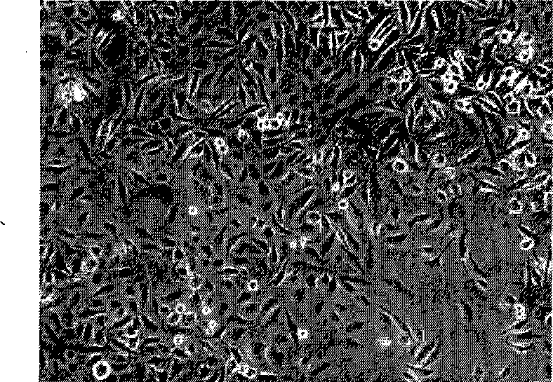
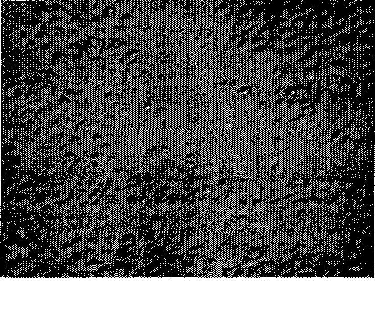
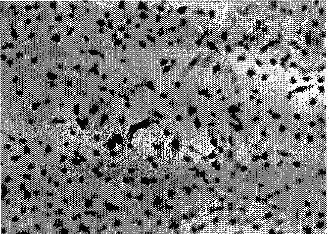
[0035] 1) Use phase contrast and differential interference inverted biological microscope to observe the growth status and shape of cells: Rca-T cells are mostly polygonal or spindle-shaped epithelio...
experiment example 3
[0048] Experimental Example 3 Subcutaneous Inoculation of Nude Mice Tumor Formation and Experimental Lung Metastasis Results
[0049] 1) Subcutaneous inoculation tumor formation experiments showed that: Rca-T and Rca-B cell lines were inoculated subcutaneously in nude mice with 3×10 6 The cells were sacrificed after 20 days, and tumors were formed in 16 subcutaneous injection points of each cell line, and the tumor formation rate was 100%. Observation of subcutaneous tumor formation tissue by pathological section of all tumor tissues showed a typical structure of squamous cell carcinoma, large cell atypia, multinucleated tumor giant cells and pathological mitosis were common, and cancer nests could be seen in the subcutaneous tumor formation tissue, which can be seen to keratinized beads (see Figure 17 ).
[0050] 2) The results of experimental lung metastasis experiment: Rca-T and Rca-B cells were inoculated into the tail vein of nude mice at 3×10 6 21 days after inoculat...
PUM
 Abstract
Description
Claims
Application Information
 - R&D
- Intellectual Property
- Life Sciences
- Materials
- Tech Scout
- Unparalleled Data Quality
- Higher Quality Content
- 60% Fewer Hallucinations
Browse by: Latest US Patents, China's latest patents, Technical Efficacy Thesaurus, Application Domain, Technology Topic, Popular Technical Reports.
© 2025 PatSnap. All rights reserved.Legal|Privacy policy|Modern Slavery Act Transparency Statement|Sitemap|About US| Contact US: help@patsnap.com


