Method for outline extraction of level set medical ultrasonic image area based on edge and statistical characteristic

A technology of ultrasonic image and statistical features, applied in the field of medical ultrasonic image processing, can solve problems such as edge blur, ultrasonic image quality is not very high, and cannot meet the actual needs of medical diagnosis

Inactive Publication Date: 2009-12-09
HARBIN INST OF TECH
View PDF0 Cites 70 Cited by
  • Summary
  • Abstract
  • Description
  • Claims
  • Application Information

AI Technical Summary

Problems solved by technology

The segmentation method based on edge detection is mainly to find the closed boundary of the region of interest, which is generally realized by various differential operators, such as Roberts operator, Sobel operator, Laplace operator and other gradient operators and wavelet transform, etc. Because the differential operation is greatly affected by noise, the quality of such methods for segmenting ultrasound images is not very high; the image segmentation method based on regional features is to divide the overall image into several complementary overlapping small areas, so that the pixels in each area A certain similarity is greater than the similarity between regions. Such segmentation methods include threshold method, region growing method, Watershed algorithm, EM algorithm, mathematical morphology method, and statistical feature method. All pixels are calculated, and the edges are often blurred, so it is not conducive to the effective extraction of edges
[0004] In recent years, on the basis of traditional pixel feature segmentation, segmentation methods based on deformation models have also been proposed, including Snakes model, optimization model, geometric and statistical feature model, physical model, random field model, etc. (M.Alemán, P.Alemán , L.Alvarez.Semiautomatic Snake-based Segmentation of Solid Breast Nodules onUltrasonography.Lectures Notes in Computer Science (EuroCast 2005), 2005, 3634: 467-472), and an efficient numerical method using level sets in solving curve evolution problems to A method for image segmentation and contour extraction (Yu-Len Huang, Yu-Ru Jiang, Dar-Ren Chen. Level Set Contouring for Breast Tumor in Sonography. Journal of Digital Imaging. 2007, 20(3): 238-247), etc. , but the segmentation effect is still not good enough
[0005] In short, for the automatic extraction of the contour of the ultrasound image area, the existing methods are not of high quality and cannot meet the actual needs of current medical diagnosis.

Method used


Image

  • Method for outline extraction of level set medical ultrasonic image area based on edge and statistical characteristic
  • Method for outline extraction of level set medical ultrasonic image area based on edge and statistical characteristic
  • Method for outline extraction of level set medical ultrasonic image area based on edge and statistical characteristic

Examples


Embodiment Construction

[0082] Below in conjunction with accompanying drawing and specific implementation example, the present invention will be further described:

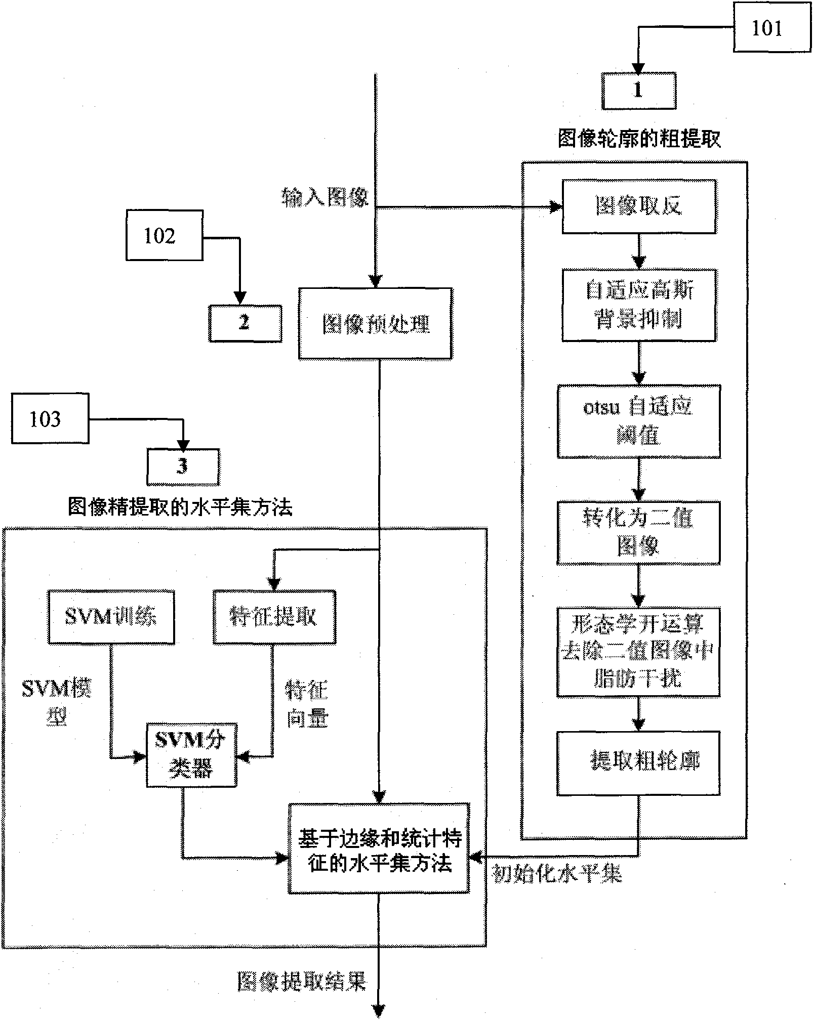
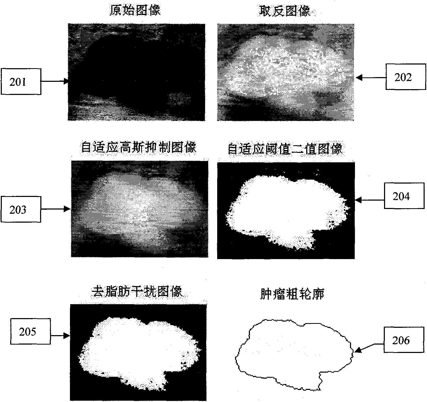
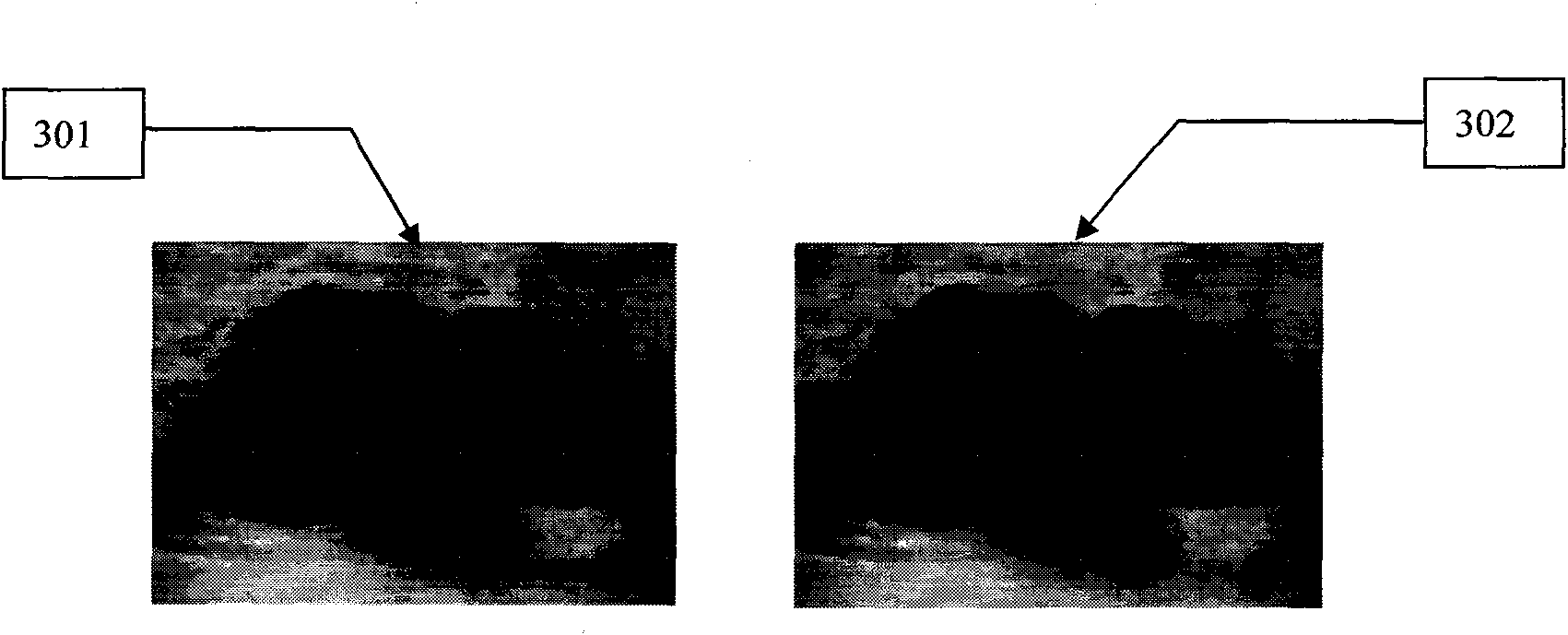
[0083]figure 1 Middle: 101 is the rough extraction of the contour of the region of interest, 102 is the ultrasonic image preprocessing based on anisotropy, and 103 is the fine extraction of the contour of the level set region based on the edge and statistical features; figure 2 Middle: 201 is the original image of breast tumor, 202 is the result of image inversion operation, 203 is the image result after applying adaptive Gaussian function, 204 is the binary image based on OTSU adaptive threshold, 205 is fat removal and other material interference After the image, 206 is the rough extraction result of the outline; image 3 Middle: 301 is the preprocessing result of the ultrasound image, 302 is the result of embedding the thick outline into the preprocessed image; Figure 4 Middle: 401 is the original breast tumor image, 402 is the leve...


PUM


Abstract

The invention provides a method for the outline automatic extraction of a level set medical ultrasonic image area based on an edge and a statistical characteristic, aiming at the characteristics of low contrast degree and small signal-to-noise ratio of medical ultrasonic images and designing a method for the outline extraction of the level set area based on the edge and the statistical characteristic. The method comprises three steps of: 1, carrying out area outline coarse extraction, completing image inverse extraction and self-adapting Gaussian function background suppression, applying otsu image automatic threshold algorithm to convert the image into a binary image, and carrying out image degreasing interference operation and closed area outline extraction work; 2, adopting a selectivity aeolotropy medical ultrasonic image smoothing algorithm to pre-treat an original image; and 3, carrying out the precise extraction of the level set image area based on the edge and the statistical characteristic. Experimental results indicate that compared with the prior method, the method of the invention can obtain a more precise partition result.

Description

(1) Technical field [0001] The invention relates to the field of medical ultrasonic image processing, in particular to a method for extracting a region outline of a medical ultrasonic image. (2) Background technology [0002] As one of the important forms of medical imaging, medical ultrasound images play an important role in clinical diagnosis and medical research. In clinical practice, the morphological features and its area or volume of the region of interest in ultrasound image diagnosis are important diagnostic information, and the calculation of these information depends on the contour of the region. Therefore, extracting the contour of the region is a key link in the diagnosis of medical ultrasound images. However, due to defects such as ultrasound signal attenuation, speckle, shadows, signal leakage, and edge loss due to the directionality of image acquisition, the boundaries between tissues in the image are blurred, which makes the doctor's attention to the outline...

Claims


Application Information

Patent Timeline
Patent Type & Authority Applications(China)
IPC IPC(8): G06T7/00
Inventor 沈毅马立勇李晓峰
Owner HARBIN INST OF TECH
Why Eureka
Try Eureka