Microfluidic chip group used for screening formyl peptide receptor agonist and screening method
A technology of microfluidic chips and formyl peptide receptors, which is applied in the direction of measuring devices, instruments, scientific instruments, etc., can solve the problems of cumbersome experimental processes, and achieve the effect of saving analysis time and reducing detection costs
- Summary
- Abstract
- Description
- Claims
- Application Information
AI Technical Summary
Problems solved by technology
Method used
Image
Examples
Embodiment 1
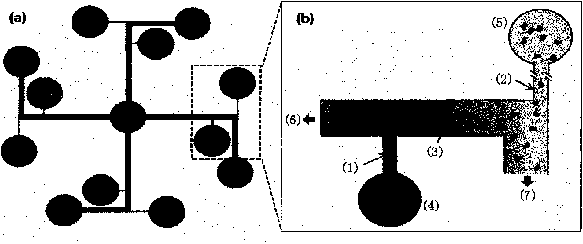
[0036] BME was taken out from the -20°C refrigerator and placed on ice. When it melted into a liquid state, pipette 0.1 μL, and figure 1The chip shown is placed under an optical microscope, and the suction head is aligned with the entrance of the basement membrane channel, and liquid BME is slowly injected into the channel. After leaving for 5 minutes, add 50% ethanol (v / v), PBS and cell culture medium to the channel of the chip in sequence, place the chip in a 37° C. incubator for 15 minutes, and the BME is completely solidified. Place the chip under an optical microscope, add 7 μL of medium to each sample pool, extract 1 μL of medium from the cell waste pool in the outer circle, and immediately inject 1 μL of cell suspension into the cell inlet, most of the cells enter the perfusion channel and stay, Only a very small number of cells enter the migration channel and place the chip in the incubator. After the cells adhered to the wall, the chip was taken out to take pictures,...
Embodiment 2
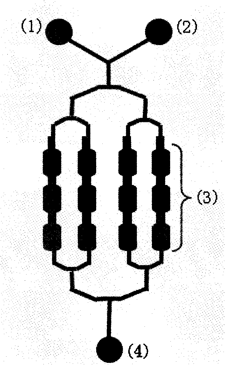
[0038] RBL-FPR cell suspension is injected from the cell inlet figure 2 For the chips shown, after the cells evenly entered each micro-culture chamber and settled, the chips were placed in the incubator overnight, and the cells fully adhered to the wall and stretched. The compound solutions (fMLF-QD and GSH-QD) labeled with quantum dots (quantum dot, QD) were introduced into the chip to stimulate the cells, and the chip was placed at 37°C CO 2 After 45 minutes in the incubator, the washing solution was introduced into the chip, and the cells were rinsed for about 45 seconds. Finally, the chip was imaged under a fluorescent microscope to detect the endocytosis of the formyl peptide receptor. The excitation wavelength of the mercury lamp was set to 330-385nm, and the detection wavelength was set to For more than 420nm, take fluorescence photos and bright field photos in the same field of view. The interaction time, concentration and washing time of compound-quantum dots and RB...
Embodiment 3
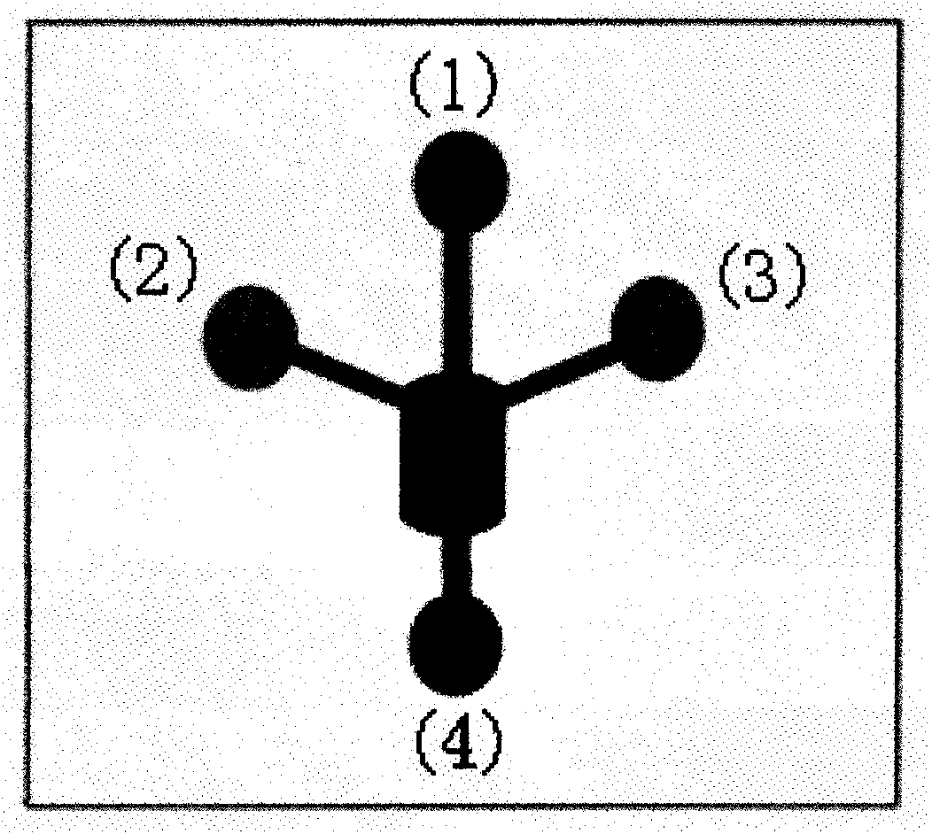
[0040] RBL-FPR cell suspension is injected from the cell inlet image 3 For the chips shown, after the cells settled, the chips were placed in the incubator overnight, and the cells fully adhered to the wall and stretched. Drain the culture medium in the sample pool, inject HBSS solution into the chip to wash the cells twice, add the calcium ion fluorescent dye Fluo-4AM diluted to 5 μg / mL with HBSS in advance, put the chip in the incubator and incubate for 45 minutes, and drain the sample pool Inject the dye in the chip, wash the cells twice by injecting HBSS solution into the chip again, and record the instantaneous flow process of calcium ions caused by the stimulation of the cells with a fluorescence microscope. Compound stimulation was applied immediately after the light source, and the CCD shutter was opened, and 50 photos were continuously taken at a speed of 1 s / frame. The effects of fMLF or GSH at the concentrations of 0nM, 4nM, 40nM and 400nM on inducing intracellula...
PUM
 Abstract
Description
Claims
Application Information
 - R&D
- Intellectual Property
- Life Sciences
- Materials
- Tech Scout
- Unparalleled Data Quality
- Higher Quality Content
- 60% Fewer Hallucinations
Browse by: Latest US Patents, China's latest patents, Technical Efficacy Thesaurus, Application Domain, Technology Topic, Popular Technical Reports.
© 2025 PatSnap. All rights reserved.Legal|Privacy policy|Modern Slavery Act Transparency Statement|Sitemap|About US| Contact US: help@patsnap.com


