Lung cancer tissue protein blotting membrane and preparation method thereof

A tissue protein and imprinting membrane technology, applied in the field of medical research, can solve the problems of cumbersome operation process, long time-consuming, limited tissue sample source, etc., and achieve the effect of improving efficiency, saving time, and efficient integration and utilization.

Inactive Publication Date: 2010-07-21
广州医学院 +1
View PDF1 Cites 10 Cited by
  • Summary
  • Abstract
  • Description
  • Claims
  • Application Information

AI Technical Summary

Problems solved by technology

In the prior art, the immunoblotting research for detecting the expression of known lung cancer proteins generally goes through the processes of clinical specimen collection, protein extraction and quantification, SDS-

Method used


Image

  • Lung cancer tissue protein blotting membrane and preparation method thereof

Examples


specific Embodiment 1

[0024] Specific Example 1: Transient receptor potential (TRPC6) channel immunoblotting

[0025] 1. Protein extraction:

[0026] (1) Cut out 100 mg of lung cancer tissue, wash the residual blood and carbon dust with pre-cooled phosphate buffered saline (PBS), dry it with filter paper, cut it into pieces, and put it into a 1-2ml glass homogenizer;

[0027] (2) For every 100 mg of lung cancer tissue, add ice-bath pre-cooled radioimmunoprecipitation analysis lysate (RIPA) and 4ul protease inhibitor-phenylmethylsulfonyl fluoride (PMSF), and homogenize on ice;

[0028] (3) Centrifuge at 10000g for 50min at 4°C;

[0029] (4) Aliquot into 0.2ml centrifuge tubes, store part at -80°C, and partly use for protein concentration determination.

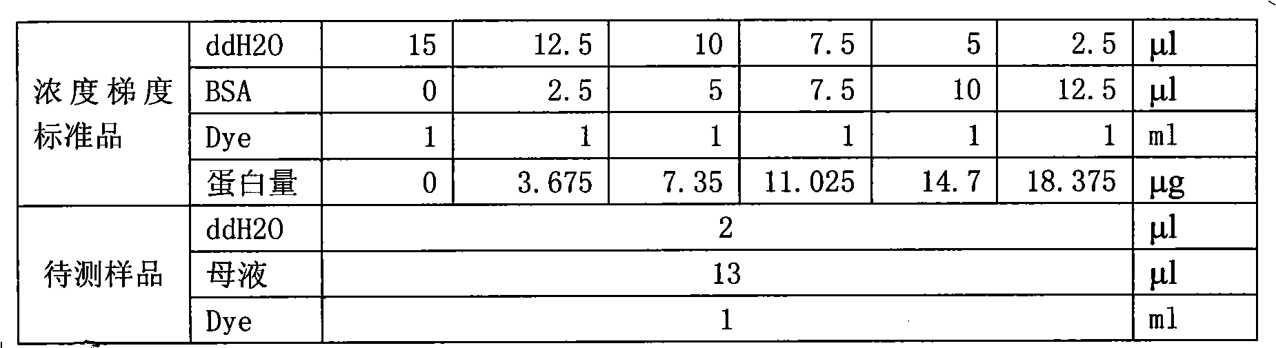
[0030] 2. Determination of protein concentration:

[0031] (1) Preparation of dye: take 8ml of dye, add 32ml of ultrapure water (1:4 dilution), mix well, filter with Wortman No. 1 filter paper, and put it on ice for later use;

[0032] (2) Prepa...

specific Embodiment 2

[0077] Specific Example 2: Cytoskeleton Protein Immunoblotting (Beta-Action)

[0078] Steps 1-6 are the same as in Example 1

[0079] 7. Immune response:

[0080] (1) Take out the PVDF membrane on the second day and add the target protein primary antibody dilution (mouse anti-Beta-Actin 1:1000, 10ul-10ml, prepared by 3% BSA-TBST; SANTA CRUZ BIOTECHNOLOGY, cat#SC-47778) , incubate in a hybridization bag shaker at 37°C for 2 hours, recover the primary antibody, add NaN3 20ul (final concentration: 0.02%), and store at 4°C;

[0081] (2) Take out the membrane, wash the membrane 3 times with TBST, 10min each time;

[0082] (3) Add target protein secondary antibody dilution (goat anti-mouse 1:7000, 2-14ml; prepared by 2.5% milk-TBST), and incubate at 37°C for 1h on a hybridization bag shaker;

[0083] (4) Take out the membrane, wash the membrane 3 times with TBST, 10 min each time.

[0084] 8. Imaging:

[0085] (1) Prepare developer and fixer in advance; prepare luminescent solu...


PUM


Abstract

The invention relates to a lung cancer tissue protein blotting membrane and a preparation method thereof. The membrane is prepared by the extraction of lung cancer tissue protein, electrophoretic separation and membrane conversion. The preparation method is characterized in that a polyvinylidene fluoride (PVDF) membrane is contained, and an instantaneous receptor potential channel TAPC6 is formed on the PVDF membrane, wherein the molecular weight is between 120kda and 85kda of the non-prestained protein molecular weight standard; or a cytoskeletal protein immune blot is formed on the activated PVDF membrane, wherein the cytoskeletal protein is above 40kda of the non-prestained protein molecular weight standard. The invention has convenient and quick storage and transportation and can be repetitively used, therefore the lung cancer specimen resource and the research are integrated and utilized efficiently; the researcher can obtain the relevant data when the subsequent immune reaction is completed after the lung cancer immune blotting, thereby saving the time of scientific research and enhancing the efficiency.

Description

technical field [0001] The invention relates to a lung cancer tissue western blot membrane and a preparation method thereof, belonging to the field of medical research. Background technique [0002] Lung cancer, also known as bronchial lung cancer, is a malignant tumor originating from bronchi and alveolar epithelial cells. Lung cancer has become the malignant tumor with the fastest growing morbidity and mortality and the greatest threat to human health and life. The 5-year survival rate of lung cancer is only 10% to 15%, and the treatment of advanced lung cancer is even more difficult. 80% of lung cancer is non-small cell lung cancer, and squamous cell carcinoma, adenocarcinoma and large cell carcinoma are the main types. Most of them are found in the middle and late stage, and the comprehensive treatment effect of surgery, radiotherapy and chemotherapy is not ideal. Additionally, 20% of lung cancers are small cell lung cancers, which have a worse overall prognosis. There...

Claims


Application Information

Patent Timeline
IPC IPC(8): G01N33/68G01N33/574G01N21/31G01N1/28B01D57/02
Inventor 何建行卢文菊张奇王健
Owner 广州医学院
Why Eureka
Try Eureka