
Tumor marker colloidal gold immunochromatographic assay quantitative detection test paper and method for preparing same

A technology of tumor markers and immunochromatography, applied in the field of colloidal gold immunochromatography quantitative detection test strips and its preparation, to achieve short detection cycle, high detection sensitivity and good specificity

Inactive Publication Date: 2010-12-01
CENT SOUTH UNIV
View PDF6 Cites 22 Cited by
  • Summary
  • Abstract
  • Description
  • Claims
  • Application Information

AI Technical Summary

Problems solved by technology

Colloidal gold immunochromatography has the advantages of simplicity, rapidity, sensitivity, and on-site operation. It has been widely used in the detection of plague, avian influenza, and pneumophila. No test strips for quantitative detection of tumor markers have been successfully developed in China. report

Method used


Image

  • Tumor marker colloidal gold immunochromatographic assay quantitative detection test paper and method for preparing same
  • Tumor marker colloidal gold immunochromatographic assay quantitative detection test paper and method for preparing same
  • Tumor marker colloidal gold immunochromatographic assay quantitative detection test paper and method for preparing same

Examples


Embodiment 1

[0028] The preparation method of test strip and test paper box of the present embodiment comprises the following steps:

[0029] 1. Antibody preparation

[0030] Use commercially available paired mouse anti-human free prostate-specific antigen (fPSA) monoclonal antibodies Ⅰ and Ⅱ (product model: A8014-1, A8014-2 Beijing Bomai Century Biotechnology Co., Ltd.), 20mM, pH7.4 Dialyze against PBS at 4°C overnight.

[0031] 2. Coating of nitrocellulose membrane

[0032] Coating buffer preparation: 0.02M phosphate buffer solution (PBS) with pH 7.4 was used as the coating buffer, which was sterilized by 0.22um microporous membrane and stored at 4°C for later use, valid for two weeks.

[0033] Preparation of blocking solution: 0.02M phosphate buffer saline (PBS) containing 0.5% BSA, pH 7.4, filtered and sterilized by a 0.22um microporous membrane, and stored at 4°C for later use, valid for one week.

[0034] Dilute the mouse anti-human free prostate specific antigen monoclonal antibo...

Embodiment 2

[0045] The usage method of the tumor marker rapid quantitative detection test paper of the present invention:

[0046] Mix the sample to be tested with the sample diluent, then insert the test paper into the sample mixture, the liquid in the sample moves upwards by siphon action, and the result is interpreted within 10-15 minutes. Quantification of tumor markers can be performed according to the following two methods:

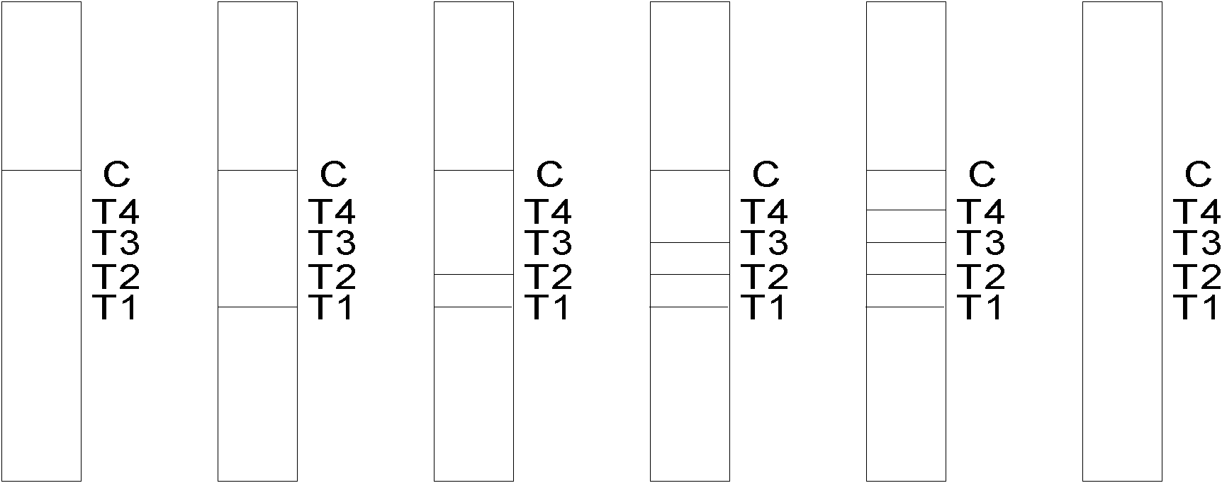
[0047] 1. Visual detection: count the number of color T-lines and observe the color depth, compare the color development table of the standard solution, and read the concentration of free PSA from the table.

[0048] Prepare free-state prostate-specific antigen colloidal gold immunochromatographic test strips according to the determined optimal conditions, and detect free-state prostate-specific antigen at different concentrations. The sample treatment solution for free-state prostate-specific antigen was prepared into 1ng / ml, 3ng / ml, 5ng / ml, 7ng / ml. Three re...

Embodiment 3

[0052] Stability test The newly prepared colloidal gold immunochromatographic test strips were stored at 37°C for 8 days, and tested with 1ng / ml, 3ng / ml, 5ng / ml, and 7ng / ml tumor marker free prostate specific antigen respectively. It shows that compared with the newly prepared detection test strip, the sensitivity is not significantly decreased, and the specificity is good.


PUM

PropertyMeasurementUnit
Widthaaaaaaaaaa
Diameteraaaaaaaaaa

Abstract

The invention discloses tumor marker colloidal gold immunochromatographic assay quantitative detection test paper and a method for preparing the same. The test paper comprises a sample pad, a gold-labeled antibody conjugate film, a nitrocellulose membrane, a water absorbing cushion and a reaction support, wherein the nitrocellulose membrane of which the end close to the sample pad is used as a starting end and the end close to the water absorbing cushion is used as a tail end is enveloped by five antibody strips in turn, namely T1, T2, T3 and T4 detection strips of mouse anti-human tumor marker monoclonal antibody I and a C quality control strip of goat anti-mouse IgG respectively; and the gold-labeled antibody conjugate film is a film coated with a colloidal gold-labeled mouse anti-human tumor marker monoclonal antibody II. The test paper and the method realize rapid and accurate quantitative detection of the tumor marker and thus meet the requirement of rapid detection of cancer surveying and screening, family cancer prognosis monitoring and the like.

Description

technical field [0001] The invention belongs to the field of biological detection, and in particular relates to a colloidal gold immunochromatographic quantitative detection test strip for tumor detection and a preparation method thereof. Background technique [0002] Tumor markers (tumor markers, TM) refer to a class of substances produced by tumor cells themselves or produced by the body's response to tumor cells during the occurrence and proliferation of tumors, reflecting the existence and growth of tumors, including Proteins, hormones, enzymes (isoenzymes) and oncogene products, etc. Some of these substances do not exist in normal human bodies and are only found in embryos, and some of them are found in tumor patients with more than normal human bodies. Tumor markers have great practical value in tumor screening, diagnosis, prognosis and prognosis, evaluation of treatment efficacy, and follow-up observation of high-risk groups. By measuring its existence or content, i...

Claims


Application Information

Patent Timeline
IPC IPC(8): G01N33/577G01N33/544
Inventor 陈真诚方成朱健铭
Owner CENT SOUTH UNIV
Learn More