Phosphorescent iridium complex capable of targeting tumor cell
A phosphorescent iridium complex, tumor cell technology, applied in fluorescence/phosphorescence, biochemical equipment and methods, microbial assay/inspection, etc., to achieve the effect of simple connection method and high photostability
- Summary
- Abstract
- Description
- Claims
- Application Information
AI Technical Summary
Problems solved by technology
Method used
Image
Examples
Embodiment 1
[0015] Embodiment 1: the preparation of iridium complex: iridium dichloro bridge complex (pq) 2 Ir(μ-Cl) 2 Ir(pq) 2 Prepared according to literature (see literature Nonoyama, K. Bull. Chem. Soc. Jpn. 1974, 47, 467-468.). Weigh IrCl 3 ·3H 2 O (5.52mmol) and the corresponding cyclometalated C^N ligand 2-phenylquinoline (11.04mmol) were added to the double-neck flask, and the mixture of 2-ethoxyethanol and water was injected with a syringe under nitrogen protection (3:1, v / v), the reaction mixture was heated to 110° C., stirred for about 24 hours, and a precipitate formed. After the reaction was stopped, the reaction mixture was cooled to room temperature, and a precipitate was obtained by filtration. The resulting precipitate was washed with water and ethanol respectively to obtain a red solid iridium dichloro bridge complex (pq) 2 Ir(μ-Cl) 2 Ir(pq) 2 . Weigh the iridium dichloro bridge complex and RGD peptide ligand with a molar ratio of 1:4 and add them into a double-...
Embodiment 2
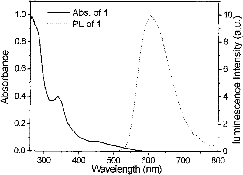
[0016] Example 2: UV-Vis absorption spectrum and phosphorescence spectrum test of the complex
[0017] The absorption and emission spectra of the complexes were measured in DMSO / MEM (1:200, v / v) solution. The complex has a strong absorption band at 280-400nm in the ultraviolet region, which can be attributed to the π-π * transition ( 1 LC's contribution. The moderately intense absorption band at 400–500 nm can be attributed to the spin-allowed charge transfer from the metal to the ligand ( 1 MLCT) transition (dπ(Ir)→π * ). In addition, a weaker absorption peak is observed above 500 nm, which can be attributed to the spin-forbidden 3 MLCT and 3 LC transition. In DMSO / MEM=1:200, the complexes all have strong emission at room temperature, and the emission wavelength is located at 610nm. The emission peak appears to be a featureless broadband emission, indicating that the emission is mainly from 3 MLCT excited state (see attached figure 1 )
Embodiment 3
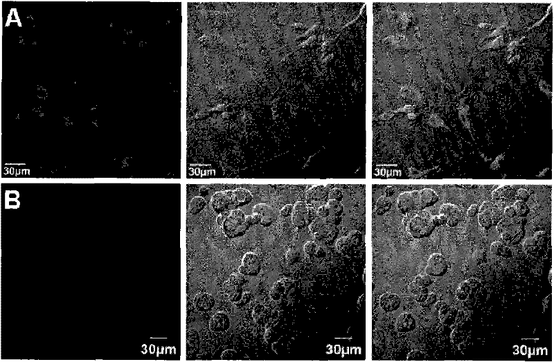
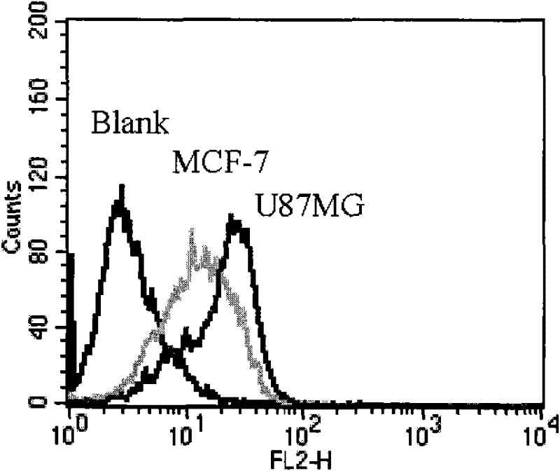
[0018] Example 3: The tumor cell imaging experiment of the complex
[0019] 1. MCF-7 (human breast cancer, integrin α v beta 3 low expression) and U87MG (human glioma, integrin α v beta 3 High expression) cell line (purchased from Shanghai Cell Bank, Chinese Academy of Sciences). MCF-7 cells were grown in MEM medium containing 10% FBS (fetal bovine serum) and 1% insulin (10 mL: 400 U), while U87MG cells were grown in MEM medium containing 10% FBS. Conditions during culture: 37°C, 5% CO 2 , saturated humidity. Replace the culture medium every two days, and subculture every 3 to 4 days.
[0020] 2. Spread the coverslips on a Ф35mm petri dish in a 1×10 5 Cells / mL seeded, 37°C, 5% CO 2 , placed in an incubator for cultivation. After the cells adhered to the wall, they were washed three times with phosphate buffered saline (PBS), and the complex solution with a final concentration of 2 μM was added to incubate at room temperature for 15 minutes, then the solution was aspir...
PUM
 Abstract
Description
Claims
Application Information
 - R&D
- Intellectual Property
- Life Sciences
- Materials
- Tech Scout
- Unparalleled Data Quality
- Higher Quality Content
- 60% Fewer Hallucinations
Browse by: Latest US Patents, China's latest patents, Technical Efficacy Thesaurus, Application Domain, Technology Topic, Popular Technical Reports.
© 2025 PatSnap. All rights reserved.Legal|Privacy policy|Modern Slavery Act Transparency Statement|Sitemap|About US| Contact US: help@patsnap.com


