

Protein suspension chip for detecting dengue antibody in serum sample and preparation method and using method thereof
A technology for suspending chips and detecting antibodies, applied in the fields of resisting vector-borne diseases, measuring devices, instruments, etc., can solve problems such as lack of models and evaluations
- Summary
- Abstract
- Description
- Claims
- Application Information
AI Technical Summary
Problems solved by technology
Method used
Image

Examples
preparation example Construction
[0043] 2. Preparation of samples to be tested
[0044] 1. Preparation of target analyte samples
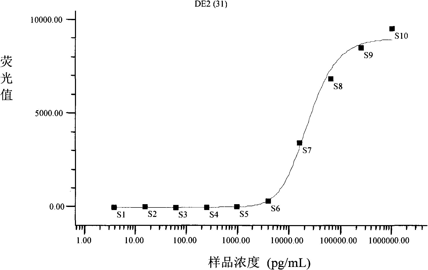
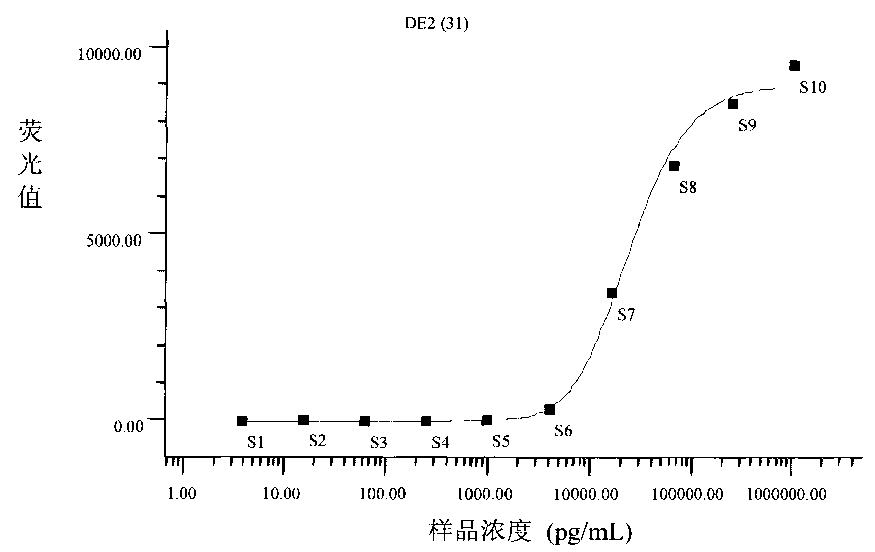
[0045] The target analyte is mouse anti-dengue fever IgG, and the interfering samples or samples used as method-specific tests are other antibodies or other proteins other than the target detection object, including rabbit anti-Tura serum, rabbit anti-avian influenza H5 serum, rabbit anti-West Nile Antibodies, BSA, casein, tryptone, etc. All the above-mentioned samples to be analyzed were dissolved in the sample diluent and stored at 4°C. The stock solution concentration of mouse anti-dengue E2 IgG was 1.06 μg / mL.
[0046] Dilute the mouse anti-dengue IgG sample diluent to be analyzed into samples of different concentrations in a 4-fold ratio to draw a standard curve of sample detection dose-response. Among them, the concentration of several samples is lower than the sensitivity of detection, and the concentration of high concentration samples The binding sites of the encoded m...
Embodiment 1
[0049] Embodiment 1, the preparation of the protein suspension chip that detects dengue fever antibody
[0050] 1. Capturing antigen-coated encoded microspheres
[0051] The coded microsphere No. 031 used in the present invention was purchased from Bio-Rad Company of the United States. The coded microsphere is used to label the dengue E2 protein antigen that can capture the dengue antibody, that is, the dengue E2 protein is used to coat the microsphere.
[0052] A. Activation of encoded microspheres
[0053] Take 100μL (1.25×10 6 pcs) encoded microspheres into a 1.5mL centrifuge tube, centrifuge at 14000×g, carefully aspirate and discard the supernatant. Add 100 μL of microsphere washing buffer to suspend, shake and sonicate, centrifuge at 14000×g, carefully aspirate and discard the supernatant. Add 100 μL of microsphere activation buffer, then add 10 μL of freshly prepared EDC (50 mg / mL), then add 10 μL of freshly prepared 50 mg / mL Sulfo-NHS, shake at high speed for 30 sec...
Embodiment 2
[0063] Embodiment 2, optimization of suspension chip preparation method conditions
[0064] 1. Selection of the amount of antigen coating on microspheres
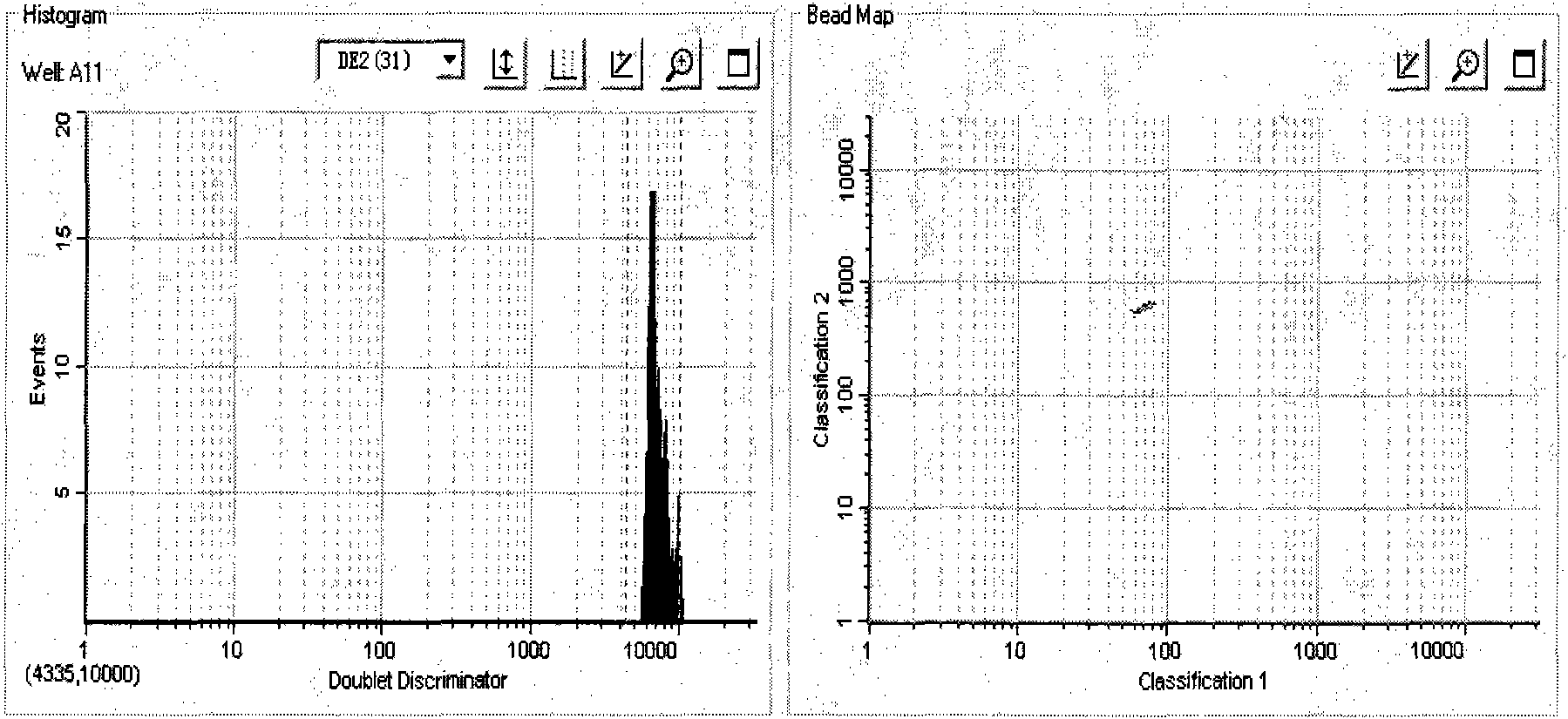
[0065] 100 μL of microspheres coded as No. 031 were coated with 1 μg, 5 μg, 10 μg, 15 μg, 20 μg, 25 μg, 30 μg, 35 μg, 40 μg, 45 μg, and 50 μg, respectively. After testing the effect comparison, with 1~50μg / 1.25×10 6 A coded microsphere, that is, 0.2-200ng / 2500-5000 microspheres / coating effect is the best. After counting under a microscope, store it in a dark place and refrigerate it for later use. Such as figure 1 As shown, the No. 031 microspheres coated with dengue E2 protein antigen all fell in the correct detection area, and obtained high signal-to-noise ratio results (MFI value was much greater than 2000), indicating that the optimized suspension chip detection system can be successfully used for dengue fever Antibody detection.
[0066] 2. Optimization of biotinylated antibodies
[0067] The present invention res...
PUM
| Property | Measurement | Unit |
|---|---|---|
| diameter | aaaaa | aaaaa |
| Sensitivity | aaaaa | aaaaa |
Abstract
Description
Claims
Application Information