
Method for preparing carcinoembryonic antigen working electrode for screen printing electrode

A technology of screen printing electrodes and working electrodes is applied in the field of preparation of carcinoembryonic antigen working electrodes, and can solve the problems of reduced detection accuracy, difficulty in removal, sensitivity and service life effects, etc.

Inactive Publication Date: 2010-12-22
NINGBO UNIV
View PDF5 Cites 10 Cited by
  • Summary
  • Abstract
  • Description
  • Claims
  • Application Information

AI Technical Summary

Problems solved by technology

[0004] However, the current amperometric sensor has the following deficiencies: 1. It is necessary to add an electronic medium (eT) such as ferrocene or potassium ferricyanide to the reaction cell for detection during use, which increases the complexity of the immunoassay system and causes the working electrode Antibodies on the surface are easily inactivated, and the sensitivity and service life are greatly affected; 2. The working electrodes are mostly used for one-time use, and the electrodes cannot be regenerated, because the immune products on the surface of the working electrodes are difficult to remove after the immune binding reaction occurs; 3. For specific detection objects with very complex backgrounds (such as serum samples), it is often necessary to undergo certain sample purification before detection, which increases the detection time and detection process and reduces the accuracy of detection

Method used


Image

  • Method for preparing carcinoembryonic antigen working electrode for screen printing electrode
  • Method for preparing carcinoembryonic antigen working electrode for screen printing electrode
  • Method for preparing carcinoembryonic antigen working electrode for screen printing electrode

Examples


Embodiment 1

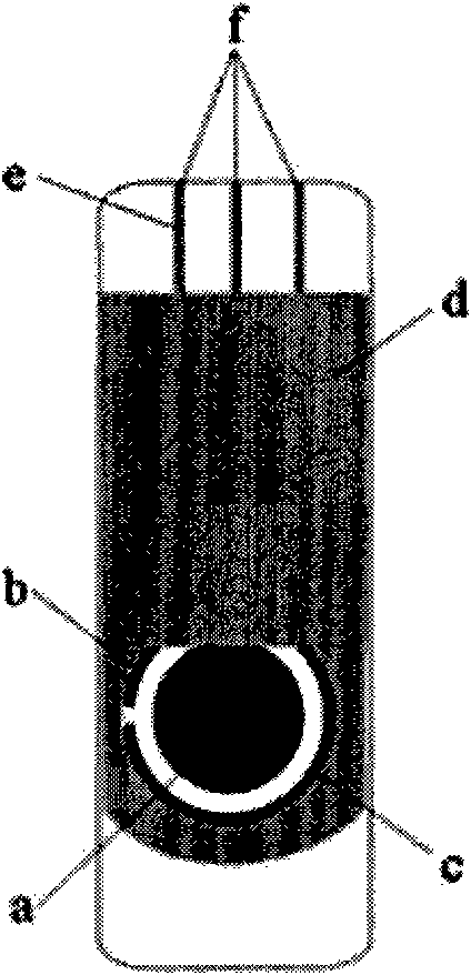
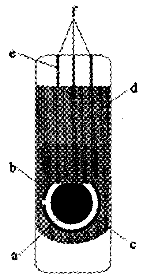
[0020] A preparation method of a carcinoembryonic antigen working electrode of a screen printing electrode, comprising the steps of:

[0021] 1) Make the concentration 0.01~0.1mol·L -1 5-10 mL of Tris-HCl buffer solution was placed in a centrifuge tube, and then added with a concentration of 1 mg·mL -1 5-10 μL of GMP solution with a diameter of 10-50 nm and a concentration of 1-3 mg·mL -1 Horseradish peroxidase-labeled mouse anti-human CEA monoclonal antibody solution 10 μL, shaking reaction at 25-37°C for 20-30 minutes;

[0022] 2) Place the above-mentioned centrifuge tube in a magnetic separator, magnetically separate it under a magnetic field of 0.1-0.3mT for 3-10 minutes, and discard the supernatant, so that the CEA antibody is coated on the surface of GMP;

[0023] 3) Add pH 7.0 to the centrifuge tube with a concentration of 0.2-1mol·L -1 5 mL of PBS buffer and a concentration of 10-15 mg·mL -1 5 μL of BSA solution, stirred at room temperature for 1 hour, magnetically...

Embodiment 2

[0028] It is basically the same as Example 1, except that the horseradish peroxidase-labeled mouse anti-human CEA monoclonal antibody is made of horseradish peroxidase-labeled goat anti-human CEA monoclonal antibody or horseradish peroxidase-labeled goat anti- Mouse CEA monoclonal antibody surrogate.


PUM

PropertyMeasurementUnit
diameteraaaaaaaaaa

Abstract

The invention discloses a method for preparing a carcinoembryonic antigen working electrode for a screen printing electrode. The method comprises the following steps of: mixing a CEA antibody with solution of gold magnetic nanoparticles, performing sealing treatment by coating the CEA antibody on the surfaces of the gold magnetic nanoparticles, sealing and fixing the coated CEA antibody, and separating the uncoated CEA antibody so as to prepare gold magnetic nanoparticle-CEA antibody solution; and forming a Nafion film on the surface of a working electrode, and adsorbing the prepared gold magnetic nanoparticle-CEA antibody on the Nafion film so as to prepare the carcinoembryonic antigen working electrode of a cation-exchange membrane of gold magnetic nanoparticle-CEA antibody-polytetrafluoroethylene. The method for preparing the carcinoembryonic antigen working electrode for the screen printing electrode has the advantages of no need of purifying the detected serum sample, no need of adding an electronic mediator during use and regeneration on the surface of the electrode after use each time.

Description

technical field [0001] The invention relates to a screen-printed electrode in an ampere immunosensor, in particular to a preparation method of a carcinoembryonic antigen working electrode of the screen-printed electrode. Background technique [0002] Carcinoembryonic antigen (CEA) is a polysaccharide-protein complex, which belongs to non-organ-specific tumor-associated antigens, and is one of the most commonly used tumor markers in tumor diagnosis and disease monitoring. Most of the tumors that secrete CEA are located in hollow organs, such as the gastrointestinal tract, respiratory tract, and urinary tract. Under normal circumstances, CEA is metabolized by the gastrointestinal tract, while CEA in the tumor state enters the blood and lymphatic circulation, causing an abnormal increase in serum CEA. For example, in patients with colorectal cancer, lung cancer, breast cancer, bladder cancer and ovarian cancer, serum CEA The amount of CEA will increase significantly, especiall...

Claims


Application Information

Patent Timeline
Patent Type & Authority Applications(China)
IPC IPC(8): G01N33/577G01N33/543G01N27/327
Inventor 姜晶晶余淑萍干宁杨欣周瑾
Owner NINGBO UNIV
Learn More