Nano-scale contrast agents and methods of use

A technology of contrast enhancers, objects, applied in the direction of applications, instruments for radiodiagnosis, general/multifunctional contrast agents, etc., which can solve the problems of lack, lack of clinical tools, limited development and effectiveness of nanosystems

Inactive Publication Date: 2011-01-19
MARVAL BIOSCI
View PDF8 Cites 2 Cited by
  • Summary
  • Abstract
  • Description
  • Claims
  • Application Information

AI Technical Summary

Problems solved by technology

Currently, the development and effectiveness of the aforementioned nanosystems is limited due to the lack of appropriate clinical tools for unambiguously and non-invasively predicting whether tumor vasculature is amenable to nanocarrier mediation in an individual, patient-specific manner. guided therapy, i.e., to determine whether a tumor has leaky vasculature
[0008] Furthermore, there is a lack of appropriate clinical tools for co-encapsulating therapeutic or anticancer agents with non-radioactive contrast enhancing agents to allow direct X-ray visualization of the biodistribution of therapeutic or anticancer agents within the body of a subject

Method used


Image

  • Nano-scale contrast agents and methods of use
  • Nano-scale contrast agents and methods of use
  • Nano-scale contrast agents and methods of use

Examples


Embodiment 1

[0043] Example 1 - Preparation and Characterization of Schematic Nanoprobes

[0044] High-concentration iodine (600 mgl / mL) was prepared by dissolving iodixanol powder (Visapaque 320 lyophilized from GE Healthcare) into ultrapure water under continuous stirring and heating at 70°C. The liposome alcohol solution was hydrated with iodine solution at 70°C, and the liposome alcohol solution contained 1,2-dipalmitoyl-sn-glycero-3-phosphocholine (DPPC), cholesterol in a molar ratio of 55:40:5 and 1,2-dipalmitoyl-sn-glycerol-3-phosphoethanolamine-N-[methoxy(poly(ethylene glycol))-2000] (Mpeg2000-DSPE), and then extruded in a LipexThermoline extruder (Northern Lipids , Vancouver, Canada) on sequential extrusion. This results in the encapsulation of the iodine solution within the central aqueous core of PEGylated liposomes. Free unencapsulated iodixanol was replaced by two days of dialysis against 300 mM NaCl using a 100,000 MWCO dialysis tubing using a saline solution (300 mM NaCl...

Embodiment 2

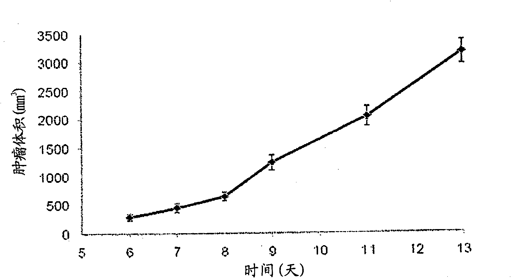
[0045] Example 2 - Imaging studies using the nanoprobes of Example 1

[0046] The nanoprobe of Example 1 was tested in a rat thoracic tumor model formed by transplanting breast cancer cells (13762MAT BIII from ATCC) into the right flank of female Fisher rats. Imaging studies started seven days after tumor implantation (tumor volume approximately 440mm 3 , see figure 1 tumor growth curve). Tumor volumes (n=15) were obtained by calipers.

[0047] Contrast-enhanced mammography was performed using a commercial digital mammography system (Senographe 2000D, GE Healthcare) at 49 kVp and 63 mAs with a rhodium target and an additional copper filter of 0.3 mm thickness. These settings are used to shape the X-rays to have an energy optimized for iodine. Under these conditions, an optimized X-ray spectrum is obtained that contains the maximum number of X-rays with energy on the k-edge of iodine, while at the same time significantly reducing the X-ray dose compared to standard mammo...

Embodiment 3

[0051] Embodiment 3-comparative embodiment (control group)

[0052] Figure 6 Whole body mammography of rats not injected with contrast agent (control group) is shown. Another control group was injected with a conventional iodinating agent (iohexol) at an iodine dose equivalent to 1,344 mgl / mL of high-dose nanoprobes. (See Figure 7 ). General vasculature and tumor lesions showed slight enhancement within the first minute after injection, but the iodinated reagent was rapidly cleared via the kidneys. (See Figure 8 )


PUM


Abstract

Compositions and methods are disclosed for evaluating a subject's vasculature integrity, for differentiating between a malignant lesion and a benign lesion, for evaluating the accessibility of a tumor to nano-sized therapeutics, for treating tumors, and for live or real time monitoring of a nano-probe's biodistribution.

Description

[0001] Cross References to Related Applications [0002] This application claims priority under 35 U.S.C. §120 to U.S. Provisional Patent Application No. 60 / 922,481, filed December 5, 2007. Additionally, this application is a continuation-in-part of U.S. Patent Application No. 11 / 595,808, filed November 10, 2006, and U.S. Patent Application No. 11 / 568,936, filed December 27, 2007, both of which were filed April 2004 Continuation-in-part of US Patent Application No. 10 / 830,190 filed on the 21st. The full text of the above-mentioned cases is hereby incorporated by reference. [0003] Statement Regarding Government-Sponsored Research or Development [0004] This invention was made with US Government support under NSF Grant No. 0401627 and NSF ERC Grant No. EEC9731643, both awarded by the National Science Foundation. The US Government has certain rights in this invention. Background technique [0005] In damaged vasculature and microvasculature, blood vessels may show incre...

Claims


Application Information

Patent Timeline
Patent Type & Authority Applications(China)
IPC IPC(8): A61B6/00
CPCA61B6/481A61B5/055A61K49/0466A61B6/504A61K49/0002A61P35/00A61P9/00
Inventor A·安娜普拉加达R·贝拉姆康达E·卡拉萨纳西斯R·M·勒波维奇
Owner MARVAL BIOSCI
Why Eureka
Try Eureka