Colloidal gold test strip for double-amplification system and preparation method thereof

A technology of colloidal gold test paper and amplification system, which is applied in biological testing, material inspection products, measuring devices, etc., can solve the problem of not further improving the sensitivity of colloidal gold, and achieve the effect of enhancing binding force, improving specificity, and improving sensitivity

Active Publication Date: 2011-06-22
GETEIN BIOTECH
4 Cites 28 Cited by

AI-Extracted Technical Summary

Problems solved by technology

[0004] Due to the sensitivity problem of colloidal gold, there i...

Abstract

The invention belongs to the field of medical clinical immediate tests, and relates to a colloidal gold test strip for a double-amplification system and a preparation method thereof. The colloidal gold test strip for the double-amplification system comprises a test strip bottom lining, and a sample pad, a polyester film coated with a gold-labeled antibody, a coating film and absorbent paper whichare lapped with and adhered to the test strip bottom lining in turn, wherein the middle part of the coating film is parallelly provided with a detection line coated with streptavidin and an F(ab)2 fragment of a monoclonal antibody of an antigen to be detected, and a control line coated with a rabbit anti-rat immunoglobulin G (IgG) antibody; the F(ab)2 fragment of the monoclonal antibody of the antigen to be detected and biotin are coated in the sample pad; and the gold-labeled antibody is colloidal gold labeled protein A and the monoclonal antibody of the antigen to be detected. The test strip has the advantages of high specificity, sensitivity and detection speed, has low cost, is easy to operate, can be widely applied to the detection of the antigen to be detected, and lays a foundationfor the timely discovery and prediction and effective detection of diseases, and an instrument and equipment are not required to be used.

Application Domain

Technology Topic

Biotin MetabolismProtein A +10

Image

  • Colloidal gold test strip for double-amplification system and preparation method thereof
  • Colloidal gold test strip for double-amplification system and preparation method thereof
  • Colloidal gold test strip for double-amplification system and preparation method thereof

Examples

  • Experimental program(2)

Example Embodiment

[0022] Example 1
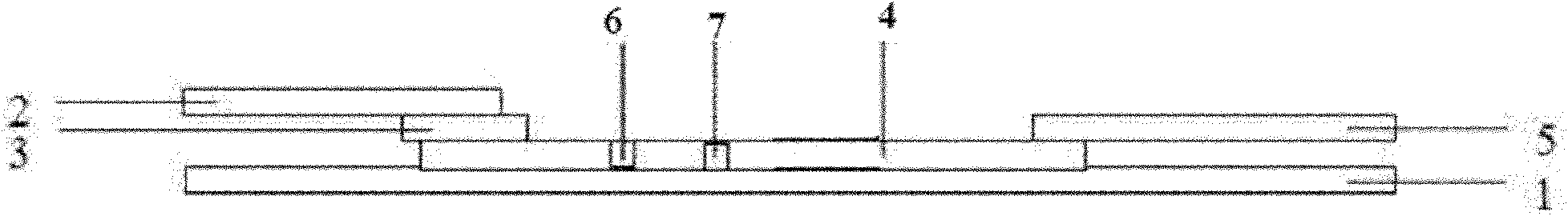
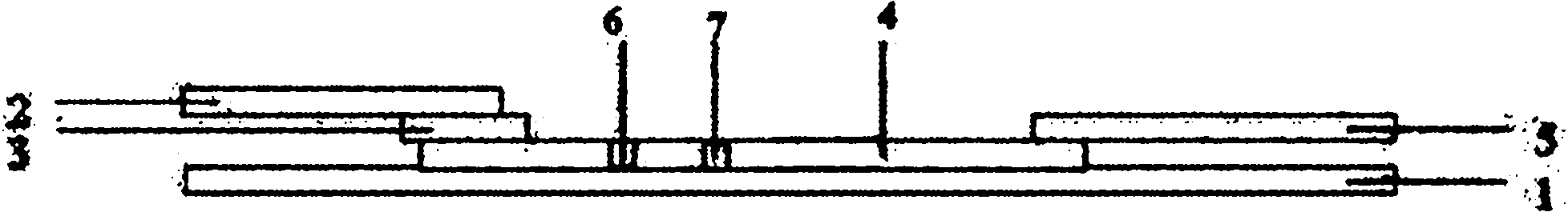
[0023] Such as figure 1 As shown, a colloidal gold test strip of a bi-amplification system includes a test strip bottom liner 1, and a sample pad 2 on the test strip bottom liner 1 that are sequentially overlapped and pasted with each other. Ester film 3, coating film 4 and absorbent paper 5, the coating film 4 is coated with a detection line 6 and a control line 7, and the middle of the coating film 4 is paralleled with a streptavidin-cardiac troponin I monoclonal antibody F(ab) of 4C2 2 Fragment detection line 6 and a control line 7 coated with rabbit anti-mouse IgG antibody, sample pad 2 is coated with F(ab) of mouse-derived cardiac troponin I monoclonal antibody 4C2 2 Fragment-Biotin, gold-labeled antibody is a gold particle labeled ProteinA, which non-specifically binds to the Fc segment of cardiac troponin I monoclonal antibody M155, and indirectly binds to the gold by the amplification of cardiac troponin I monoclonal antibody M155 On the particles. The above streptavidin, 4C2 monoclonal antibody, M155 monoclonal antibody, and rabbit anti-mouse IgG antibody were all purchased from Fitzgerald.
[0024] The specific preparation method of colloidal gold test strips is as follows:
[0025] Preparation of coating film:
[0026] Preparation of coating buffer: filter 0.025M, pH7.4 PBS with a 0.22u membrane, and place it at 4°C for use. The validity period is 7 days.
[0027] Preparation of blocking solution: filter PBS containing 1% BSA, 1% sucrose, 0.025M, pH 7.5 with 0.22 U membrane, and place it at 4°C for use. The validity period is 3 days.
[0028] Cardiac Troponin I monoclonal antibody 4C2 F(ab) 2 Fragment preparation: The monoclonal antibody 4C2 was digested with trypsin, and the Fc fragment was removed by SPA-Sepharose CL-4B gel chromatography column, and F(ab) was collected 2 Fragment.
[0029] Streptavidin-F(ab) 2 Fragment preparation: combining streptavidin and monoclonal antibody 4C2 F(ab) 2 The fragment is chemically coupled by glutaraldehyde, and then purified by affinity chromatography.
[0030] Streptavidin-F(ab) 2 Preparation of fragment detection line 6: the antibody F(ab) 2 The fragments were dried at a concentration of 4mg/ml, a peristaltic pump delivery volume of 0.4ml/min, a scribing speed of 50m/20min, and air blast drying at 20°C in a drying box for 12 hours.
[0031] Preparation of control line 7: The rabbit anti-mouse IgG antibody was 8mg/ml in concentration, the peristaltic pump delivery volume was 0.4ml/min, the scribing speed was 50m/20min, and it was air-dried in a drying box at 20°C for 12 hours.
[0032] Preparation of coating film 4: The nitrocellulose membrane containing the detection line 6 and the control line 7 was sealed at 37°C for 60 minutes with the above-mentioned sealing solution, and then dried at 37°C for two hours after being taken out, and the bag was sealed for use.
[0033] Preparation of polyester film 3 coated with gold-labeled antibody:
[0034] Preparation of chloroauric acid aqueous solution: Dissolve 10g of chloroauric acid in 1000ml of tri-distilled water, prepare a 1% aqueous solution, and place it at 4°C for use. The validity period is 3 months.
[0035] Preparation of trisodium citrate: Dissolve trisodium citrate with three-distilled water, make a 1% aqueous solution, filter with 0.22U membrane, and place at 4°C for use. The validity is 7 days.
[0036] Preparation of 1% potassium carbonate aqueous solution: Dissolve 1g of potassium carbonate in 100ml with three-distilled water, filter with 0.22U membrane, and place at 4°C for use. The validity period is 7 days.
[0037] Preparation of gold-labeled antibody preservation solution: Dissolve 15g of sucrose and 20ul of sodium azide in 100ml of 1% BSA with pH=7.4, filter with 0.22U membrane, and place at 4°C for use. The validity period is 7 days.
[0038] Preparation of colloidal gold particles: dilute 1% chloroauric acid to 0.01% with tri-distilled water, place at 95°C for 10 minutes, add 1ml of trisodium citrate, continue the reaction for 15 minutes, wait until the color of the colloidal gold solution changes from blue After the purple turns red, 2ml of 1% potassium carbonate solution is added after cooling. The appearance should be pure, translucent, free of precipitation and floating matter.
[0039] Preparation of gold-labeled antibody: adjust the pH value of colloidal gold to 7.5 with 1% potassium carbonate solution, add ProteinA to 20 micrograms of ProteinA/ml colloidal gold, mix and react for 15 minutes, centrifuge at 10000rpm for 10 minutes, and remove the supernatant. The precipitate was blocked and washed twice with 1% BSA, and the supernatant was removed for the last time. The precipitate was dissolved with one-tenth of 1% pH=7.4 BSA, and then 5ug antibody/ml colloidal gold was added to cardiac troponin I. Clone the antibody, mix and react for 15 minutes, wash twice with the labeling washing solution, and then dissolve it with the gold-labeled antibody storage solution. Keep it at 4°C for use, the validity period is 7 days.
[0040] Spread the marked colloidal gold evenly on the polyester film with a spray volume of 0.9ul/cm*3, dry, seal the bag, and set aside.
[0041] Processing of the sample pad: soak the sample pad in 100mM PBS buffer for several hours, take it out and dry, and press 20ug-F(ab) of mouse-derived cardiac troponin I monoclonal antibody 4C2 2 Fragment-Biotin/Amount of each sample pad (22mm*300mm) Spray mouse-derived cardiac troponin I monoclonal antibody 4C2 F(ab) 2 Fragment-Biotin.
[0042] The backing 1, the sample pad 2 and the absorbent paper 5 are common components in this field. The sample pad 2, the polyester film 3 coated with gold-labeled antibody, the coating film 4, and the absorbent paper 5 are sequentially lapped and pasted to each other to obtain a test board, and finally the test board is cut into test strips of different widths.
[0043] The application of the aforementioned colloidal gold test strips in the detection of cardiac troponin I.
[0044] This test strip can be used to detect a liquid sample of cardiac troponin I. During operation, drop whole blood, plasma or serum on the sample pad 2 of this test strip. When the result appears on the test strip When a purple-red band appears on the test line 6 and the control line 7 respectively, it is a positive result, indicating that the sample contains cardiac troponin I; when only a purple-red band appears on the control line 7 of the test strip, A negative result, indicating that the sample contains cardiac troponin I or the concentration of cardiac troponin I contained in the sample is below the detection limit of this test strip; there is no purple-red band at the control 8 position of the test strip When the result is invalid.

Example Embodiment

[0045] Example 2 The specific preparation method of the colloidal gold test strip using the conventional method is as follows:
[0046] Cardiac troponin I monoclonal antibodies 4C2 and M155 (pairing antibodies) were purchased from Fitzgerald.
[0047] Preparation of coating film:
[0048] 1) Preparation of coating buffer: filter 0.025M, pH7.4 PBS with a 0.22u membrane, and place it at 4°C for use. The validity period is 7 days.
[0049] 2) Preparation of blocking solution: filter PBS containing 1% BSA, 1% sucrose, 0.025M, and pH 7.5 with a 0.22U membrane, and place it at 4°C for use. The validity period is 3 days.
[0050] 3) F(ab) of monoclonal antibody 4C2 2 Preparation of the segment: the monoclonal antibody was digested with trypsin, passed through the SPA-Sepharose CL-4B gel chromatography column to remove the Fc fragment, and collected F(ab) 2 Fragment.
[0051] 4)F(ab) 2 The preparation of section detection line 6: The antibody is at a concentration of 4mg/ml, the peristaltic pump delivery volume is 0.4ml/min, the scribing speed is 50m/20min, and air-dried in a drying box at 20°C for 12 hours.
[0052] 5) Preparation of control line 7: The rabbit anti-mouse IgG antibody is 8mg/ml in the concentration, the peristaltic pump delivery volume is 0.4ml/min, the scribing speed is 50m/20min, and it is air-dried for 12 hours at 20°C in a drying box.
[0053] 6) Preparation of coating film 4: The nitrocellulose NC film containing the detection line and the control line was sealed at 37°C for 60 minutes with the above-mentioned sealing solution, and then dried at 37°C for two hours after being taken out, and the bag was sealed for use.
[0054] Preparation of polyester film coated with gold-labeled antibody:
[0055] 1) Preparation of chloroauric acid aqueous solution: Dissolve 10 g of chloroauric acid in 1000 ml of tri-distilled water, prepare a 1% aqueous solution, and place it at 4°C for use, with a validity period of 3 months.
[0056] 2) Preparation of trisodium citrate: Dissolve trisodium citrate with three-distilled water, prepare a 1% aqueous solution, filter with 0.22U membrane, and place it at 4°C for use. The validity period is 7 days.
[0057] 3) Preparation of 1% potassium carbonate aqueous solution: Dissolve 1 g of potassium carbonate in 100 ml with three-distilled water, filter with 0.22U membrane, and place it at 4°C for use. The validity period is 7 days.
[0058] 4) Preparation of gold-labeled antibody preservation solution: Dissolve 15g of sucrose, 20ul of sodium azide in 100ml of 1% BSA with pH=7.4, filter with 0.22U membrane, and place at 4℃ for standby, the validity period is 7 days .
[0059] 5) Preparation of colloidal gold particles: Dilute 1% chloroauric acid to 0.01% with tri-distilled water, place it at 95°C for 10 minutes, add 1ml of trisodium citrate, continue the reaction for 15 minutes, wait until the color of the colloidal gold solution changes After the blue turns from purple to red, 2ml of 1% potassium carbonate solution is added for use after cooling. The appearance should be pure, translucent, free of precipitation and floating matter.
[0060] 6) Preparation of gold-labeled antibody: adjust the pH value of colloidal gold to 7.5 with 1% potassium carbonate solution, add 20 μg monoclonal antibody M155/ml colloidal gold, mix and react for 15 minutes, centrifuge at 10000rpm for 10 minutes, The supernatant and the precipitate were blocked and washed twice with 1% BSA, washed twice with the labeling washing solution, and then dissolved in the gold-labeled antibody preservation solution, placed at 4°C for use, and the validity period was 7 days.
[0061] 7) Spread the marked colloidal gold evenly on the polyester film with a spray volume of 0.9ul/cm*3, dry, seal the bag, and set aside.
[0062] The backing 1, the sample pad 2 and the absorbent paper 5 are common components in this field. The sample pad 2, the polyester film 3 coated with gold-labeled antibody, the coating film 4, and the absorbent paper 5 are sequentially lapped and pasted to each other to obtain a test board, and finally the test board is cut into test strips of different widths.
[0063] Comparison of the sensitivity and specificity of the two methods
[0064]
[0065] Remarks: "+" indicates a positive result, the number of "+" indicates the degree of coloration, that is, the severity of the positive, the more the color, the higher the severity of the positive, the "-" indicates that the color is not developed, that is, the negative result.
[0066] It can be seen from the above table that the colloidal gold test strips of the two large systems of the present invention can significantly improve the detection sensitivity and specificity.
[0067] The monoclonal antibody of the test antigen of the present invention is not limited to the cardiac troponin I monoclonal antibodies 4C2 and M155 used in the examples, and any antibody or antigen that can bind to the test antigen or antibody can be used In the present invention, the double amplification system. In addition, the present invention is not limited to the application in the field of colloidal gold, and can also be used in the immune response in the field of fluorescence and latex.

PUM

no PUM

Description & Claims & Application Information

We can also present the details of the Description, Claims and Application information to help users get a comprehensive understanding of the technical details of the patent, such as background art, summary of invention, brief description of drawings, description of embodiments, and other original content. On the other hand, users can also determine the specific scope of protection of the technology through the list of claims; as well as understand the changes in the life cycle of the technology with the presentation of the patent timeline. Login to view more.

Similar technology patents

High-temperature resistant aluminum powder paint

Owner:QINGDAO CREEK NEW ENVIRONMENTAL PROTECTION MATERIAL

Classification and recommendation of technical efficacy words

  • Improve binding
  • High sensitivity

Fc variants with altered binding to FcRn

ActiveUS20060173170A1Improve bindingLong retentionImmunoglobulinsFermentationImmunology
Owner:XENCOR

System and method for optical coherence imaging

ActiveUS20060055936A1Sufficient signal to noise ratioHigh sensitivityInterferometersScattering properties measurementsPhysicsCoherent imaging
Owner:THE GENERAL HOSPITAL CORP

Catheter-free implantable needle biosensor

ActiveUS20080033269A1High sensitivitySimple stepSurgeryVaccination/ovulation diagnosticsNeedle electrodeElectricity
Owner:VIRTUE MEDICAL TECH (HUZHOU) CO LTD
Why Eureka
Try Eureka