Real time in situ characterization method for single biomolecular reaction

A biomolecular and reaction technology, applied in biological testing, individual particle analysis, particle and sedimentation analysis, etc.

Inactive Publication Date: 2011-11-23
SHANGHAI INST OF APPLIED PHYSICS - CHINESE ACAD OF SCI
View PDF2 Cites 7 Cited by
  • Summary
  • Abstract
  • Description
  • Claims
  • Application Information

AI Technical Summary

Problems solved by technology

[0004] However, there is no real-time in situ imaging method for single...

Method used


Image

  • Real time in situ characterization method for single biomolecular reaction
  • Real time in situ characterization method for single biomolecular reaction
  • Real time in situ characterization method for single biomolecular reaction

Examples


Embodiment 1

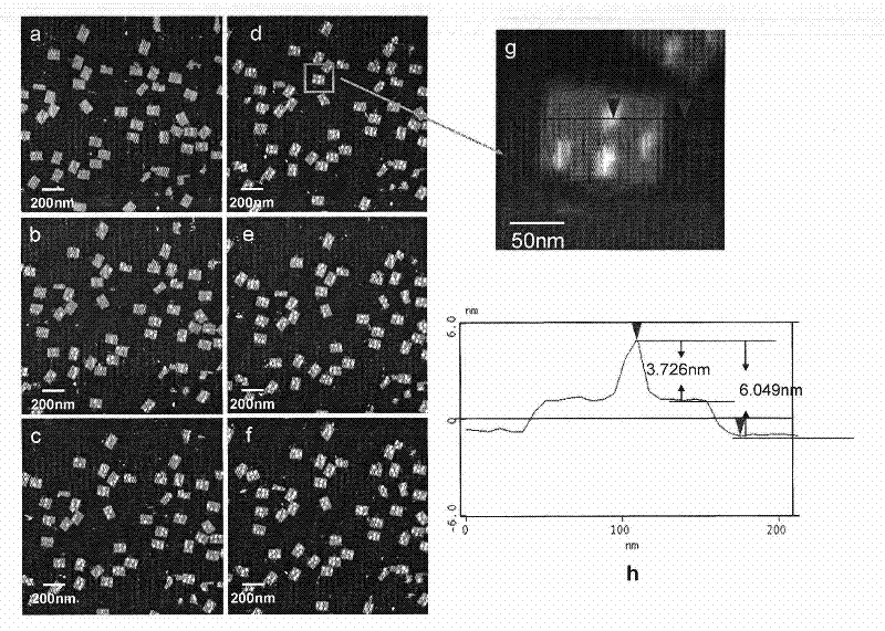
[0035] 1. The DNA origami designed in this example utilizes single-strand self-assembly of M13mp18 DNA and staples to form a rectangular pattern with a length and width of 100nm and 70nm, respectively, such asfigure 1 As shown, at four positions on the DNA origami, the ends of the staple strands are biotinylated for binding to streptavidin.

[0036] Preparation of specific biotinylated square DNA origami:

[0037] M13mp18 DNA single strands and staple single strands (including biotinylated staple single strands) were mixed at a molar concentration of 1:10 and placed in 1×TAE / Mg 2+ Buffer system (Tris 40mM, acetic acid 20mM, EDTA 2mM, MgCl 2 12.5mM, and the pH value is 8), the total volume is 61uL, and then placed on the PCR instrument and annealed from 95°C to 20°C at an annealing speed of 0.1°C / 10s, and stored at 4°C after the reaction is completed.

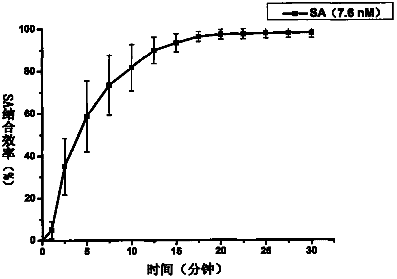
[0038] 2. Real-time in situ detection of biotin dynamic reaction process between streptavidin and biotin-modified DNA origam...


PUM


Abstract

The invention discloses a real time in situ characterization method for a single biomolecular reaction. The method comprises the following steps: immobilizing a reactant I on a DNA origami design site; then carrying out atomic force microscope imaging for the reactant I-modified DNA origami; adding a reactant II, wherein the reactant II is correspondingly combined with the reactant I, meanwhile continuously scanning imaging and collecting data. With the method provided by the present invention, the real time in situ detection can be performed, the single biomolecular reaction process can be recorded exactly. In addition, the method is benefit for studying the dynamic process of the single biomolecular reaction on the interface.

Description

technical field [0001] The invention relates to a real-time in-situ characterization method for a single biomolecule reaction. Background technique [0002] So far, most single-molecule studies still use the method of optical microscopy, the labeling step is inevitable, and the fluorescence lifetime must also be considered. At the same time, according to the characteristics of single-molecule research, it is necessary to fix some components in the detection system and carefully adjust the capture molecules and detection molecules to obtain a high signal-to-noise ratio; in addition, the non-specific adsorption that plagues the detection is also special consideration must be given. On the other hand, the force spectroscopy method based on atomic force microscopy (AFM) can give a quantitative description of the interaction force between molecules, and the accuracy can reach the PN level, which is one of the important methods for studying single molecules; however, the detectio...

Claims


Application Information

Patent Timeline
IPC IPC(8): G01N15/10G01N33/50G01N33/68
Inventor 吴娜李宾胡钧
Owner SHANGHAI INST OF APPLIED PHYSICS - CHINESE ACAD OF SCI
Why Eureka
Try Eureka