Flow measurement system for detecting tumor cells and analysis and monitoring method

A tumor cell and flow measurement technology, applied in biochemical equipment and methods, material stimulation analysis, microbial measurement/testing, etc., can solve complex time-consuming procedures, error-prone analysis speed, time-consuming and other problems, and achieve fast and accurate classification Effect

Inactive Publication Date: 2014-03-12
TIANJIN UNIV +1
View PDF5 Cites 2 Cited by
  • Summary
  • Abstract
  • Description
  • Claims
  • Application Information

AI Technical Summary

Problems solved by technology

However, the method of identifying cells using light microscopy has limitations and is difficult to use in the rapid analysis of enriched blood samples containing very small numbers of circulating tumor cells for the following reasons:
First, commonly used optical microscopes (such as fluorescence microscopes, bright-field or dark-field microscopes, etc.) are designed based on the principle of non-diffraction imaging, and their images are formed by two-dimensional projection of the three-dimensional structure of cells. The measured cell structure features are structural two-dimensional projection features, which cannot truly reflect the three-dimensional structural shape and characteristics of cells
Second, since the microscopic image is a two-dimensional projection of the three-dimensional structure of the cell, it usually requires very complex image analysis methods to identify cells based on the image analysis, especially when analyzing cells with complex three-dimensional structure and morphology, which generally requires manual analysis , so the image analysis method based on the optical microscope is difficult to automate, and the related optical microscope operation and image measurement also need manual operation, which is time-consuming, error-prone and extremely low analysis speed
Third, immunofluorescence staining often requires expensive reagents and complex and time-consuming procedures
Due to these problems, although most of the current commercial flow cytometers use laser beams as excitation beams, they cannot distinguish cells by measuring and analyzing diffraction images.

Method used


Image

  • Flow measurement system for detecting tumor cells and analysis and monitoring method
  • Flow measurement system for detecting tumor cells and analysis and monitoring method
  • Flow measurement system for detecting tumor cells and analysis and monitoring method

Examples


Embodiment Construction

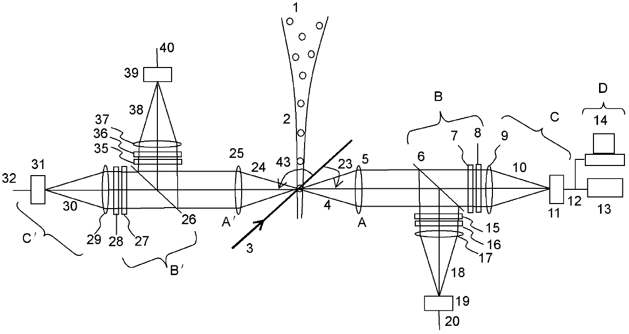
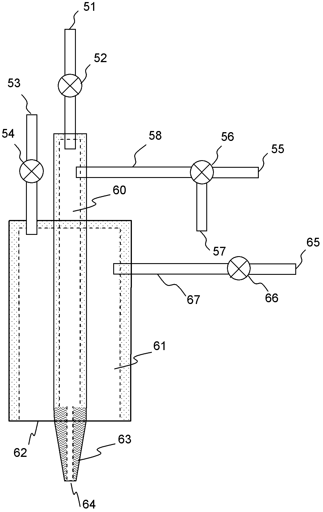
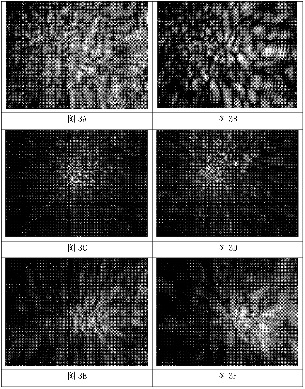
[0086] The flow measurement system for detecting tumor cells and the analysis and monitoring method of the present invention will be described in detail below with reference to the embodiments and accompanying drawings.

[0087]The flow measurement system for detecting tumor cells and the analysis and monitoring method of the present invention use an adjustable imaging system to measure diffraction images of different wavelengths and polarizations, and then use computer software to quickly analyze the statistical parameter vectors of multiple diffraction image modes and compare these parameter vectors with The cell type discrimination criterion can quickly classify a large number of cells to achieve the purpose of detecting tumor cells. The method described in the present invention can also be realized by other optical systems, methods and computer software. The specific method is to use filtration or centrifugation to remove the small cells in the blood sample whose linear si...


PUM


Abstract

The invention discloses a flow measurement system for detecting tumor cells and an analysis and monitoring method. The system comprises a sample steam formed by enriched cells, a sample stream chamber for storing the sample stream, a sheath tube for storing a sheath fluid, a coherent excitation light beam intersecting with the sample stream, a scattered light receiving objective lens portion for measuring a coherent scattering light beam having a central scattering angle from excited cells, a light splitting and filtering portion, an imaging measurement and data output portion, an image processing circuit, a computer portion and a display portion. The method comprises the following steps of: preparing an enriched cell suspension from a blood or lymph sample, generating coherent scattering light signals, collecting corresponding diffraction image data of different wavelengths and polarizations and transmitting the data into the image storage and processing unit in the computer, extracting image pattern characteristics, determining the positions of the detected cells in the diffraction image characteristic parameter vector sample space, and displaying and outputting the corresponding statistical analysis results. The system and method can rapidly and accurately classify a large amount of cells to achieve the purpose of detecting tumor cells.

Description

technical field [0001] The present invention relates to a flow measurement system and analysis method for detecting tumor cells, in particular to a diffraction image with adjustable wavelength and polarization formed by measuring coherently scattered light of tumor cells present in peripheral blood samples and other cell samples of patients by flow means Signal, calculating and extracting the image features highly correlated with the three-dimensional structure features inside the cells, which can automatically, quickly and accurately identify tumor cells and provide their distribution data for the flow measurement system and analysis and monitoring methods for detecting tumor cells. Background technique [0002] Researchers have found that malignant solid tumors originating from human epithelial tissue are the most prevalent cancers, such as lung, breast, prostate, pancreatic, and colorectal cancers. Malignant solid tumors will generate their own microvascular system during...

Claims


Application Information

Patent Timeline
Patent Type & Authority Patents(China)
IPC IPC(8): G01N21/63C12Q1/02
Inventor 冯远明胡新华
Owner TIANJIN UNIV
Why Eureka
Try Eureka