Epidermal differentiation microRNA signature and uses thereof

An epidermis, expression-level technology, applied in the field of skin, can solve problems such as expensive equipment, inconvenient operation, and multiple follow-up steps

Active Publication Date: 2012-08-01
LOREAL SA
View PDF5 Cites 13 Cited by
  • Summary
  • Abstract
  • Description
  • Claims
  • Application Information

AI Technical Summary

Problems solved by technology

This also requires extensive use of two-dimensional electrophoresis gel technology, so it is very inconvenient to operate
In addition, accurate ...

Method used


Image

  • Epidermal differentiation microRNA signature and uses thereof
  • Epidermal differentiation microRNA signature and uses thereof
  • Epidermal differentiation microRNA signature and uses thereof

Examples


example 1

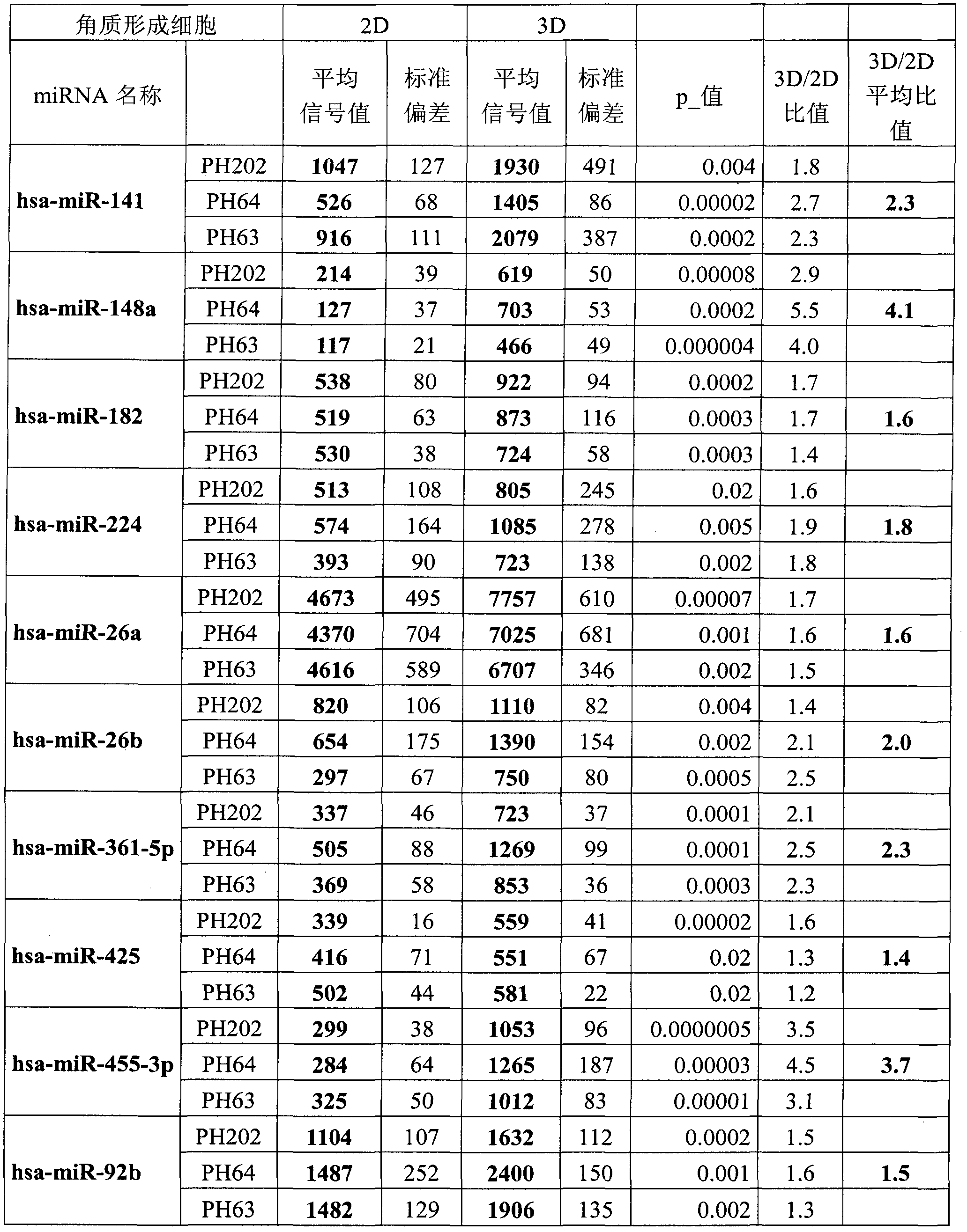
[0196] Example 1: The listed 10 miRNAs are overexpressed in differentiated regenerating epidermal keratinocytes compared to undifferentiated monolayer cultured keratinocytes.

[0197] Three primary normal human keratinocyte cell lines (PH202, PH64, and PH63) derived from mammoplasty were used in this study. The expression of all known human miRNAs was quantified in cultured keratinocyte monolayers, ie undifferentiated keratinocytes (2D) and simultaneously in these same keratinocytes in regenerated epidermal samples (3D).

[0198] The expression of each miRNA was compared statistically between the "keratinocytes in culture" sample and the "regenerated epidermal keratinocytes" sample, and compared using a Student's t-test giving the probability p. For each cell line studied, the expression level of each miRNA listed in Table 1 in the epidermis was statistically significantly higher than that in undifferentiated keratinocytes (p<0.05). For each cell line, the expression ratio 3D...

example 2

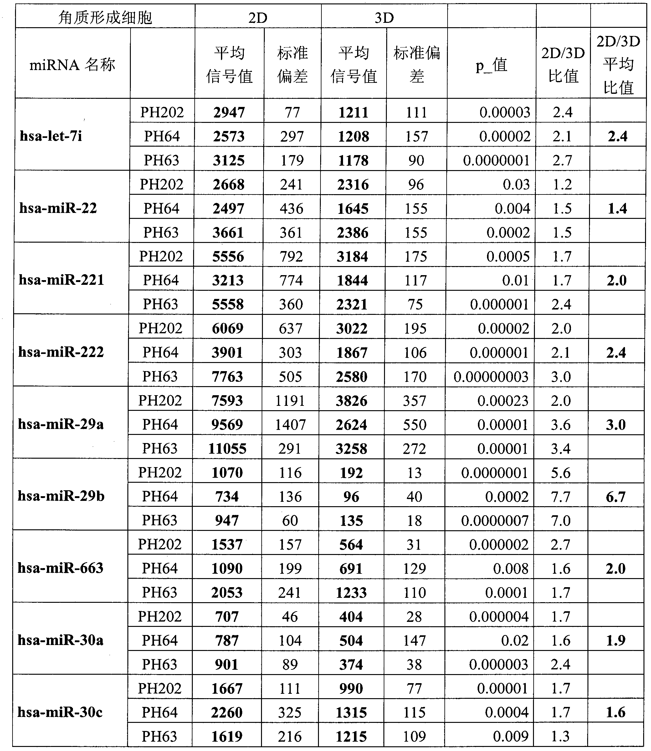
[0201] Example 2: Nine miRNAs listed are overexpressed in undifferentiated monolayer cultured keratinocytes compared to differentiated regenerating epidermal keratinocytes.

[0202]Three primary normal human keratinocyte cell lines (PH202, PH64, and PH63) derived from mammoplasty were used in this study. The expression of all known human miRNAs was quantified in cultured keratinocyte monolayers, ie undifferentiated keratinocytes (2D) and simultaneously in these same keratinocytes in regenerated epidermal samples (3D). The expression of each miRNA was statistically compared between the "cultured keratinocytes" sample and the "regenerated epidermal keratinocytes" sample, and compared using a Student's t-test for which probability p was obtained. For each cell line studied, the expression levels of each miRNA listed in Table 1 were statistically higher in the epidermis than in undifferentiated keratinocytes (p<0.05). The expression ratio 3D / 2D is given for each cell line. For e...

example 3

[0205] Example 3: Method for detecting miRNAs.

[0206] Northern Blot:

[0207] Qualitative and quantitative using conventional methods. Multiple miRNAs can be analyzed in the same experiment. This method requires the use of 5-50 μg of total RNA.

[0208] RT Q-PCR

[0209] This method can study the precursors of miRNAs and is suitable for studying mature miRNAs. This method is commercially available (TaqMan MicroRNA Assays from Applied Biosystems) and is described in several articles [23].

[0210] miRNA Microarray Analysis (Microarray)

[0211] This method is similar to conventional mRNA microarray analysis, and is based on fluorescent labeling of complementary cDNAs of miRNAs in samples, and immobilizing these labeled cDNAs, hybridizing on slides loaded with immobilized miRNA sequences [25].

[0212] Some authors argue that this miRNA microarray technology is an option for profiling the global expression profile of miRNAs if the novel sequences of miRNAs derived ...


PUM


Abstract

The present invention relates to a method for determining the state of epidermal differentiation of a human keratinocyte in a human epithelium sample, said method including determining the level of expression of at least one microRNA selected from among hsa-miR-455-3p, hsa-miR-141, hsa-miR-148a, hsa-miR-182, hsa-miR-224, hsa-miR-26a, hsa-miR-26b, hsa-miR-361-5p, hsa-miR-425, hsa-miR-92b, hsa-let-7i, hsa-miR-22, hsa-miR-221, hsa-miR-222, hsa-miR-29a, hsa-miR-29b, hsa-miR-663, hsa-miR-30a, and hsa-miR-30c within said keratinocyte, and comparing the level of expression of the microRNA(s) selected at the mean level of expression of the microRNA(s) selected in the undifferentiated keratinocytes or in differentiated epidermal keratinocytes, preferably from the same strain or the same population. The present invention also relates to various uses of the method for determining the state of epidermal differentiation, as well as to pharmaceutical or cosmetic compositions.

Description

technical field [0001] The present invention relates to cosmetics and is suitable for application to the skin. More specifically, the present invention relates to the characterization of the epidermal differentiation state of the skin, and the treatment of lesions, diseases or pathologies associated with skin differentiation. Background technique [0002] The epidermis is composed of multiple layers of epithelial cells that act as a protective barrier against the environment and stimuli. It is mainly composed of keratinocytes, which proliferate in the deepest layer of the epidermis, then begin to differentiate, and migrate from the deep layer of the epidermis to the surface. On the surface of the epidermis, most of the differentiated keratinocytes, known as keratinocytes, form the stratum corneum, which is highly protective. The epidermis is thus the site of both cellular proliferation that occurs within the basal layer and cellular differentiation in the middle layer, cul...

Claims


Application Information

Patent Timeline
IPC IPC(8): C12Q1/68A61K8/72A61Q19/00
CPCC12Q2600/178C12Q1/6881C12Q1/6883A61Q19/00A61Q19/007C12Q2600/158A61Q19/08A61K8/606A61P17/00A61P17/02A61P17/06A61P37/08A61K8/72A61Q19/005C12Q2525/207
Inventor 克莱尔·马里奥内弗朗索瓦丝·贝尔内
Owner LOREAL SA
Why Eureka
Try Eureka