Method for extracting residual nucleic acid from HE (haematoxylin eosin) dyeing piece

A technique for dyeing slides and nucleic acids, applied in the field of nucleic acid extraction, to achieve good practicality, reduce degradation, good economic benefits and social effects

Active Publication Date: 2012-09-19
JIANGSU PROVINCIAL HOSPITAL OF TCM +2
View PDF14 Cites 6 Cited by
  • Summary
  • Abstract
  • Description
  • Claims
  • Application Information

AI Technical Summary

Problems solved by technology

The above-mentioned technologies and processes are all aimed at samples such as fresh tissue cells, blood or paraffin tissue, and do not involve the extraction and...

Method used


Image

  • Method for extracting residual nucleic acid from HE (haematoxylin eosin) dyeing piece
  • Method for extracting residual nucleic acid from HE (haematoxylin eosin) dyeing piece
  • Method for extracting residual nucleic acid from HE (haematoxylin eosin) dyeing piece

Examples


Embodiment 1

[0024] The decolouring method of embodiment 1 HE staining sheet

[0025] Immerse the HE-stained slide in xylene for 30 minutes, then gently slide off the cover glass. If the cover glass cannot slide off automatically, you can extend the time appropriately; take out the cover glass and continue soaking the HE-stained slide for 8-12 hours to fully remove the neutral gum; Soak in new xylene for 15 minutes; take out the cell sheet and soak in absolute ethanol for 10-20 minutes to fully remove xylene; replace with new absolute ethanol, soak for 15 minutes, then soak in 95% ethanol for 10 minutes; soak in 80% ethanol for 5 minutes ; Soak in 70% ethanol for 2 minutes; wash with tap water for 2 minutes; decolorize with 1% hydrochloric acid alcohol hematoxylin, and control it under the microscope until the cell sheet is almost colorless, which takes about 10 minutes. ; Soak the cell sheet in 2% oxalic acid aqueous solution for 3-5 minutes. If there is still color or the original sheet ...

Embodiment 2

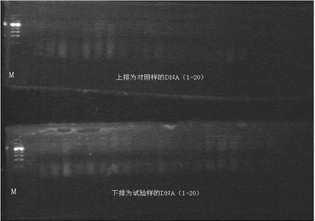
[0028] Select 20 cases of HE-stained slides from 2008 to 2011 (gastroscopic specimens, provided by the Department of Pathology, Jiangsu Provincial Hospital of Traditional Chinese Medicine), one for each case, 20 test samples were decolorized by the decolorization process in Example 1, enriched to obtain target cells, and then used E.Z.N.A. from OMEGA bio-tekTM The Tissue DNA Kit kit was used to extract DNA from the enriched target cells, and the QIAGEN RNeasy? FFPE Kit kit was used to extract RNA from the enriched tumor tissue. The other 20 control samples were directly used without the decolorization step in Example 1. Corresponding extraction kit for DNA / RNA extraction, the results are as follows figure 1 As shown in Table 1, the DNA electrophoresis band extracted after decolorization is more complete than the DNA electrophoresis band extracted from tissue without decolorization. In the figure, the upper row is the DNA of the control sample (without decolorization step), The...

Embodiment 3

[0034] One piece of HE-stained slide after conventional processing of the puncture sample was used as the material (provided by the Pathology Department of Jiangsu Provincial Hospital of Traditional Chinese Medicine in 2009 for routine puncture pathological HE-stained slide) for decolorization, and the steps were as follows:

[0035] Immerse a puncture HE sample in xylene for 30 minutes, and gently slide off the cover glass; take out the cover glass, continue to soak the HE stained piece for 8~12 hours, and fully remove the neutral gum; replace it with new xylene, and soak for 15 minutes; Soak in water and ethanol for 10-20 minutes to fully remove xylene; replace with new anhydrous ethanol, soak for 15 minutes, then soak in 95% ethanol for 10 minutes; 80% ethanol for 5 minutes; 70% ethanol for 2 minutes; tap water for 2 minutes Decolorization with 1% hydrochloric acid alcohol hematoxylin takes about 10 minutes; soak in 2% oxalic acid aqueous solution for 20 minutes; wash with t...


PUM


Abstract

The invention discloses a method for extracting residual nucleic acid from an HE (haematoxylin eosin) dyeing piece, and the method comprises the following steps of: decoloring the HE dyeing piece by using low concentration hydrochloric acid alcohol as a decoloring agent first, then, bleaching by an oxalate solution, and then extracting nucleic acid by enriching target cells. Compared with the prior art, the method provided by the invention can be used for effectively removing basic dye haematoxylin and acidic dye eosin, reducing the degradation of nucleic acid in the extracting and storing process effectively, and enriching and detecting needed target cells so as to extract residual nucleic acid by using all regular HE dyeing pieces such as a puncture tissue, a cell smear (including sputum and urea), gastrointestinal endoscope and the like and perform subsequent molecular neuropathologic detection, so that the method provided by the invention is good in practicality, and can be used for achieving good economical benefit and social benefit.

Description

technical field [0001] The invention relates to a method for extracting nucleic acid, in particular to a method for extracting residual micronucleic acid from HE stained slices. Background technique [0002] HE staining is a commonly used pathological slide preparation method, which is widely used in pathological diagnosis and research. HE staining uses two biological dyes, the basic dye hematoxylin and the acid dye eosin, to act on the nucleus and cytoplasm respectively, so that the fine structure of the cell changes its refractive index through color, so that it can be clearly presented under the light microscope The cell image shows that there are acidic substances and basic substances in the tissue cells. The acidic substances of the cells are combined with the cations of the basic dyes, and the basic substances of the nuclei are combined with the anions of the acidic dyes, so that the acidic nuclei are stained with basic hematoxylin. The alkaline cytoplasm is stained r...

Claims


Application Information

Patent Timeline
IPC IPC(8): G01N1/28
Inventor 赖仁胜谢玲陈劼吴晓斌徐慧李惠孙怡郑燕影赵苏苏赵明
Owner JIANGSU PROVINCIAL HOSPITAL OF TCM
Why Eureka
Try Eureka