Electrochemical miRNA (micro Ribose Nucleic Acid) detection method based on DNA (Deoxyribose Nucleic Acid) three-dimensional nano structure probe

A three-dimensional nanometer and detection method technology, applied in biochemical equipment and methods, microbial determination/inspection, etc., can solve problems such as a large number of target miRNAs, and achieve the effects of low cost, high stability and strong specific selectivity

Active Publication Date: 2013-01-30
SHANGHAI INST OF APPLIED PHYSICS - CHINESE ACAD OF SCI
View PDF2 Cites 27 Cited by
  • Summary
  • Abstract
  • Description
  • Claims
  • Application Information

AI Technical Summary

Problems solved by technology

[0007] The purpose of the present invention is to provide a method of ultrasensitive electrochemical miRNA using DNA three-dimensional

Method used


Image

  • Electrochemical miRNA (micro Ribose Nucleic Acid) detection method based on DNA (Deoxyribose Nucleic Acid) three-dimensional nano structure probe
  • Electrochemical miRNA (micro Ribose Nucleic Acid) detection method based on DNA (Deoxyribose Nucleic Acid) three-dimensional nano structure probe
  • Electrochemical miRNA (micro Ribose Nucleic Acid) detection method based on DNA (Deoxyribose Nucleic Acid) three-dimensional nano structure probe

Examples


Embodiment 1

[0033] Reagents include:

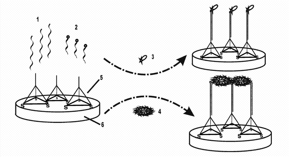
[0034] Four single-stranded DNAs assembled to form a tetrahedral probe with a three-dimensional nanostructure of DNA, Tetra-A (75bp, molecular weight 23071.0, ssDNA), Tetra-B (55bp, molecular weight 17018.0, 5' end modified sulfhydryl ssDNA), Tetra-C (55bp, molecular weight 16898.0, ssDNA modified with thiol at the 5' end), Tetra-D (55bp, molecular weight 16877.0, ssDNA modified with thiol at the 5' end), both were purchased from Dalian Takara Biological Co., Ltd. The four single-stranded DNAs contain three structural domains, each of which is complementary to the corresponding domains of the other three single-stranded DNAs (17 pairs of bases are complementary), and each single-stranded DNA surrounds one face of the tetrahedral structure. Circle, containing two bases (non-complementary, flexible) at each vertex for bending, the 3' and 5' ends of single-stranded DNA converge at the four vertices of the tetrahedron, and Tetra-A extends at the 5' end ...

Embodiment 2

[0069] The avidin-modified horseradish peroxidase (avidin-HRP) was replaced by polyavidin-modified horseradish peroxidase (poly-HRP80), and other reagents and DNA were the same as in Example 1. Both poly-HRP80 and poly-HRP80 dilutions were purchased from Fitzgerald Industries International. poly-HRP80 is a supramolecular enzyme complex containing 400 (80*5) HRP molecules, while avidin-HRP only contains about 20 HRP molecules.

[0070] The experimental procedure was the same as in Example 1, except that the avidin-modified horseradish peroxidase was replaced with poly-avidin-modified horseradish peroxidase (poly-HRP80), and other parameters remained unchanged.

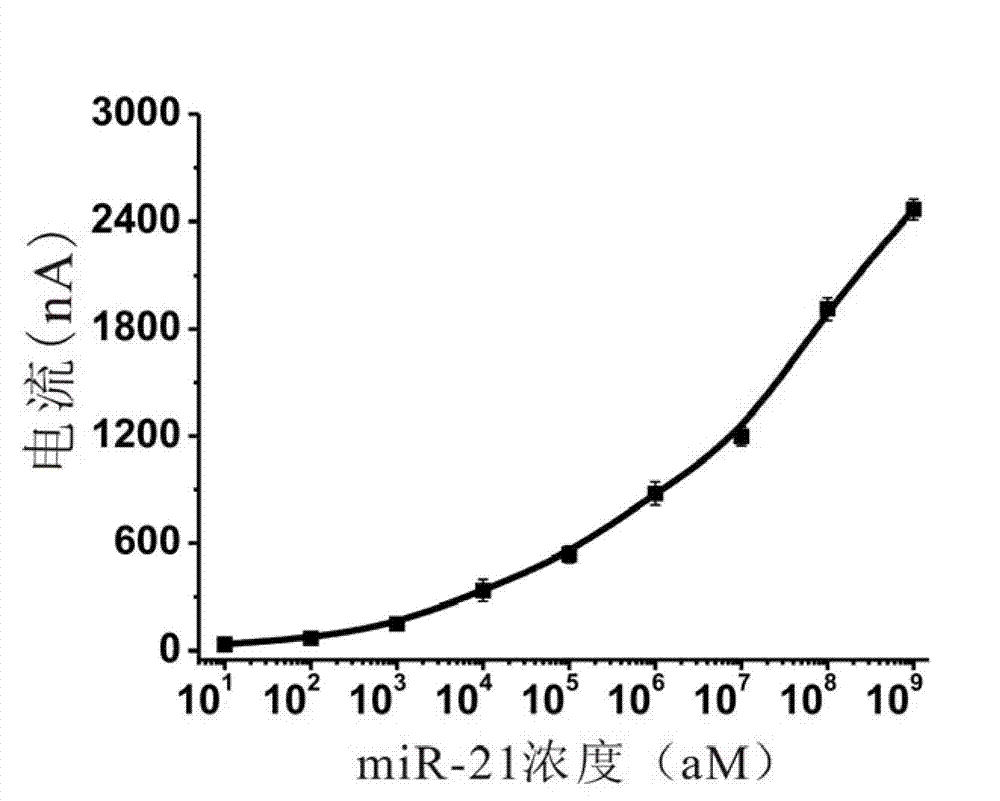
[0071] The result is as image 3 As shown, the use of polyavidin-modified horseradish peroxidase (poly-HRP80) can greatly improve the detection sensitivity. From the graph we can see that the method using tetrahedral probes according to the present invention can detect miRNAs as low as 10 aM.

Embodiment 3

[0073] Replace the Tetra-A used in the self-assembled tetrahedral probe in Example 1 with Tetra-NA (55bp, molecular weight 16959.0, ssDNA), the DNA sequence of other synthesized tetrahedral probes is the same as in Example 1, and the synthesis method is also the same , thus assembling to form a tetrahedral DNA without a recognition sequence. The sequence is as follows:

[0074] Tetra-NA (SEQ ID NO:8): 5'-ACA TTC CTA AGT CTG AAA CAT TACAGC TTG CTA CAC GAG AAG AGC CGC CAT AGT A-3' (without recognition sequence)

[0075] The first tetrahedron probe and the second tetrahedron probe without recognition sequence were synthesized according to the method in Example 1. The concentrations of the first and second tetrahedral probes were both 1 μM, and mixed according to different ratios, and then 3 μL of the two tetrahedral probe mixtures were co-assembled on the electrode surface, and left overnight at room temperature. In this way, the density of the first tetrahedron probe on the el...


PUM

PropertyMeasurementUnit
Diameteraaaaaaaaaa

Abstract

The invention provides an electrochemical miRNA (micro Ribose Nucleic Acid) detection method based on a DNA (Deoxyribose Nucleic Acid) three-dimensional nano structure probe. The electrochemical miRNA detection method comprises the steps of: synthesizing a DNA three-dimensional nano structure probe through a self-assembly method, wherein the DNA three-dimensional nano structure probe comprises one section of extended recognition sequence; assembling the DNA three-dimensional nano structure probe on the surface of a working electrode of an electrochemical device; hybridizing a target miRNA with the DNA three-dimensional nano structure probe on the surface of the working electrode; and adding oxidordeuctase and a corresponding substrate, and carrying out electrochemical detection by using the electrochemical device. The method can be used for detecting the miRNA of 10aM, therefore, the problem of the great demand on test samples in the detection method in the prior art is solved. In addition, the method has strong specificity selection and can be well used for distinguishing base pair mismatching of same family of miRNA. Compared with the method using the single-chain DNA probe, the electrochemical miRNA detection method is higher in stability.

Description

technical field [0001] The invention belongs to the field of nucleic acid hybridization detection, and in particular relates to an electrochemical miRNA detection method based on a DNA three-dimensional nanostructure probe. Background technique [0002] MicroRNAs (miRNAs) are a newly discovered class of endogenous non-coding single-stranded small RNAs with a length of 19-23 nucleotides, which usually regulate cell proliferation, differentiation, apoptosis, metabolism and other processes at the post-transcriptional level . Since Lee et al. first discovered lin-4 in Caenorhabditis elegans in 1993, which is involved in the regulation of timing development in nematodes, people have become very interested in studying the important role of these small molecules in the basic life processes of animals and plants. A large number of studies have shown that abnormal miRNAs expression levels are directly related to various types of human cancers (Nature Reviews Cancer, 2006, 6, 259-269...

Claims


Application Information

Patent Timeline
IPC IPC(8): C12Q1/68
Inventor 樊春海闻艳丽林美华
Owner SHANGHAI INST OF APPLIED PHYSICS - CHINESE ACAD OF SCI
Why Eureka
Try Eureka