Establishment method of diabetic vasculopathy model based on microfluidic chip

A technology of microfluidic chips and blood vessels for diabetes, which is applied in the measurement/testing of microorganisms, biochemical equipment and methods, artificial cell constructs, etc., and can solve the problem of monocytes being in a blank stage.

Inactive Publication Date: 2013-03-20
DALIAN INST OF CHEM PHYSICS CHINESE ACAD OF SCI
View PDF4 Cites 5 Cited by
  • Summary
  • Abstract
  • Description
  • Claims
  • Application Information

AI Technical Summary

Problems solved by technology

At present, the simulation of tissue engineering using microfluidic chips, especially the simulation of diabetic vascular lesions and the research and analysis of the movement characteristics of monocytes on the surface of the lesion are still in a blank stage.

Method used


Image

  • Establishment method of diabetic vasculopathy model based on microfluidic chip
  • Establishment method of diabetic vasculopathy model based on microfluidic chip
  • Establishment method of diabetic vasculopathy model based on microfluidic chip

Examples


Embodiment 1

[0029] Establishment of Diabetic Vascular Lesion Model Using HUVEC Cells

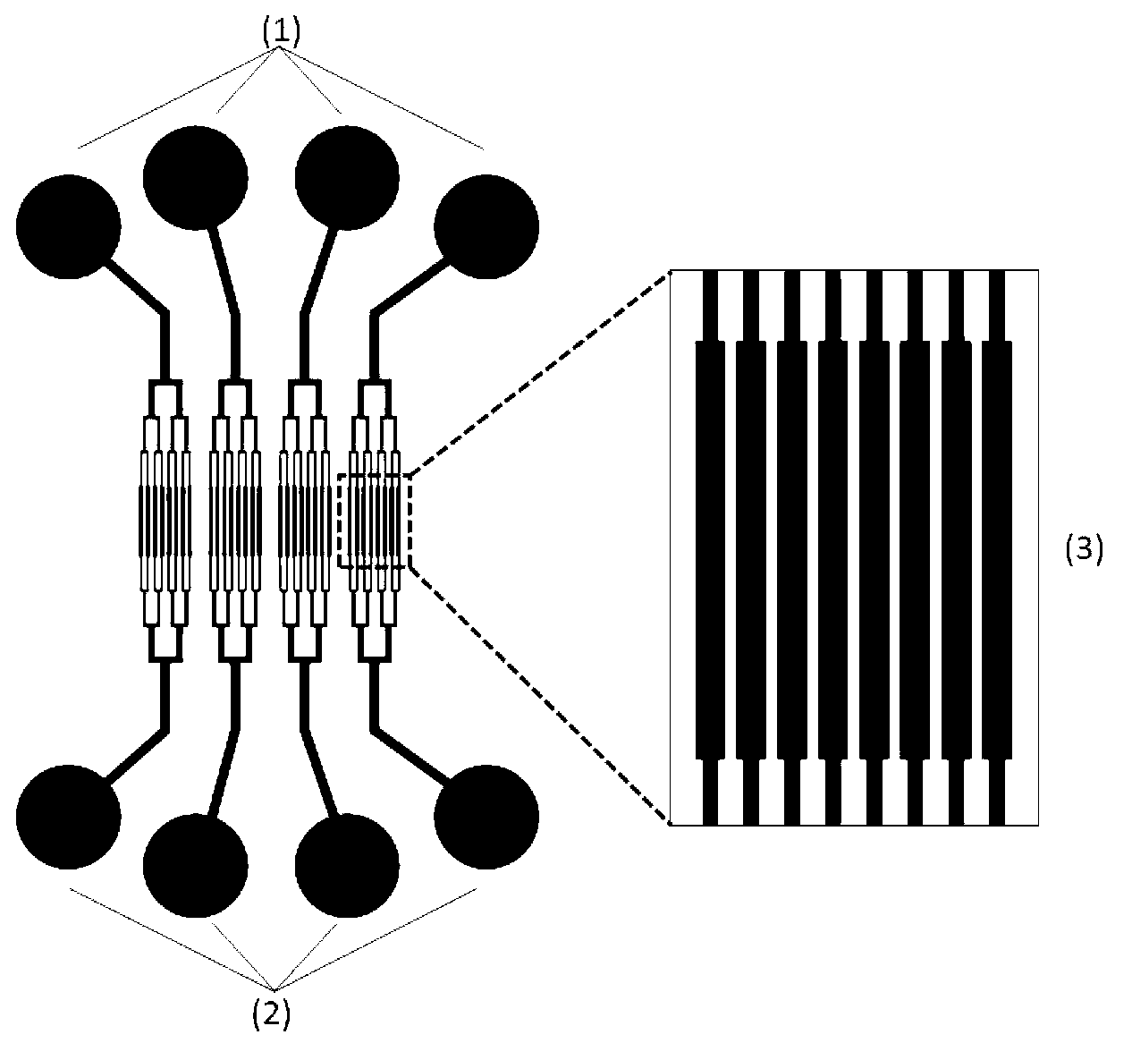
[0030] Using the microfluidic chip designed and manufactured by the laboratory, the structure is as follows: figure 1 shown. Prepare a type I mouse tail collagen working solution with a concentration of 100 μg / mL, inject it into the chip through the cell inlet pool, let it stand at room temperature overnight, remove the collagen working solution after 24 hours, and wash the channel 3 times with the cell culture solution. After digesting HUVEC cells, dilute to a concentration of 5×10 6 cells / mL cell suspension, add 100 μL HUVEC cell suspension to the chip through the cell inlet pool, under the modification of collagen, the cells quickly adhere to the wall and spread evenly on the bottom of the cell culture chamber, when the cells are observed under an optical microscope When evenly distributed in the cell culture chamber, immediately move the chips into the CO2 incubator to continue culturing. After t...

Embodiment 2

[0032]Characterization of corresponding structural and functional proteins in cells and surfaces of diabetic vascular disease model

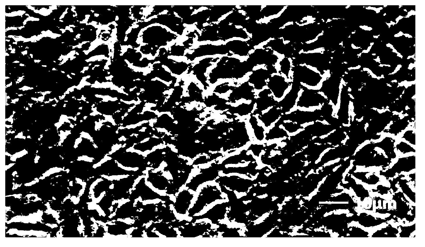
[0033] Using the microfluidic chip designed and manufactured by the laboratory, the structure is as follows: figure 1 shown. After chip modification, the same cell inoculation and culture methods as in Example 1 were used to establish a diabetic vascular disease model. After stimulation for 48 hours, immunofluorescence staining was performed on the cells, and the monitored proteins were carbon monoxide synthase (eNOS), von Willebrand factor (vWF), integrin β4 (Intβ4), vinculin, and endothelial cell adhesion molecule- 1 (VCAM-1) and intercellular adhesion molecule-1 (ICAM-1). The method is as follows: cells were fixed with 4% paraformaldehyde, washed three times with PBS buffer, 10 min each time; treated with 0.1% triton X-100 porogen for 10 min, washed three times with PBS buffer, 10 min each time; blocked with goat serum for 1 h, Primary ant...

Embodiment 3

[0035] Adhesion and rolling of monocytes on the surface of endothelial cells under different fluid shear forces

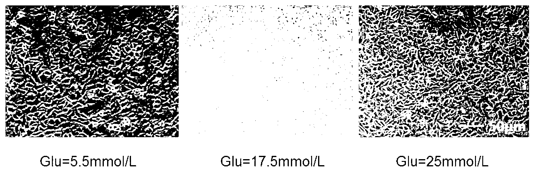
[0036] Using the microfluidic chip designed and manufactured by the laboratory, the structure is as follows: figure 1 shown. After chip modification, the same cell inoculation and culture methods as in Example 1 were used to establish a diabetic vascular disease model. Then the leukemia mononuclear cells cultured to a certain density were diluted into a certain density of cell suspension, the concentration was 1×10 6 cells / mL, through the cell inlet pool at a certain flow rate into the channel stimulated by the culture solution containing different glucose concentrations, the microscope continuously photographed the adhesion and rolling phenomenon of monocytes on the surface of HUVEC cells, and counted the occurrence of adhesion and rolling The number of monocytes was analyzed by Image ProPlus software to analyze the speed and track of monocyte movement, and the ...


PUM


Abstract

An establishment method of a diabetic vasculopathy model based on a microfluidic chip is especially directed at vascular wall adhesiveness characteristic variation resulted from vascular endothelial inflammation in diabetic vasculopathy process, and phenomena of adhesion and rolling of mononuclear cells on surfaces of endothelial cells, thrombosis and vascular obstruction under simulation of blood flow conditions. The microfluidic chip is composed of a cell inlet pool (1), a cell culture chamber (2) and an outlet pool (3), wherein the upper of the cell culture chamber (2) is connected with the cell inlet pool (1), and the lower of the cell culture chamber (2) is connected with the outlet pool (3). The establishment method can integrate vasculopathy simulation under the condition of high blood sugar and interaction studies of mononuclear cells and lesion endothelial cell surfaces under the blood flow conditions in one, and can simultaneously realize simulation of the lesion site and in-situ simulation of thrombosis in complications of diabetes.

Description

technical field [0001] The present invention relates to the technical field of applying microfluidic chip technology to tissue bionics and real-time monitoring of cell biology research, in particular to a method for establishing a model of diabetic vascular disease based on a microfluidic chip, which is applied to monocytes in A method for studying the motion characteristics of lesion surfaces. Background technique [0002] Medical research shows that 50% of cardiovascular and cerebrovascular diseases are caused by diabetes. The increased prevalence of vascular disease in the diabetic population is mainly due to the accelerated rate of atherosclerosis. Compared with non-diabetic individuals, individuals with diabetes or impaired glucose tolerance are often accompanied by other metabolic abnormalities that promote atherosclerosis; at the same time, diabetes increases the procoagulant state and accelerates the formation of atherosclerosis. Thus, patients with diabetes develo...

Claims


Application Information

Patent Timeline
Patent Type & Authority Applications(China)
IPC IPC(8): C12M3/00C12N5/071C12Q1/02
Inventor 秦建华许慧马慧朋
Owner DALIAN INST OF CHEM PHYSICS CHINESE ACAD OF SCI
Why Eureka
Try Eureka