

Gene probe composition and kit for acute lymphocytic leukemia detection
A technology of acute lymphocytes and gene probes, applied in the direction of recombinant DNA technology, microbial measurement/inspection, biochemical equipment and methods, etc., can solve problems such as deletion of heterozygosity
- Summary
- Abstract
- Description
- Claims
- Application Information
AI Technical Summary
Problems solved by technology
Method used
Image

Examples
Embodiment 1
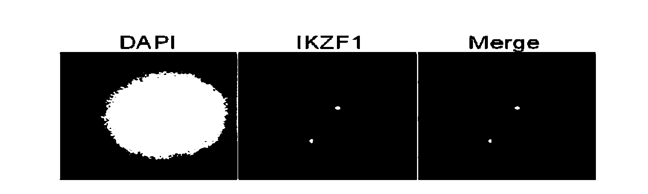
[0072] The present invention will be further described below through specific examples, but protection scope of the present invention is not limited. Embodiment 1: The preparation method of IKZF1 gene probe comprises the following steps:
[0073] 1. Primer design and clone screening: The IKZF1 gene is located in the 12.3-12.2 segment of the short arm of human chromosome 7 (7p12.3-p12.2), and all clones containing the IKZF1 gene were searched in the UCSC genome browser, NCBI Clone Registry, and Ensembl Genome Browser databases , and the optimal clone containing the gene was screened by polymerase chain reaction, numbered RP11-663L2 (as shown in Table 2).
[0074]2. Cloning culture and identification: purchase clone RP11-663L2 (Invitrogen, USA), take 8 microliters of clone bacteria solution and add it to 5 milliliters of LB liquid medium containing chloramphenicol resistance, shake and activate for 12 hours at 37°C ; Then add all the bacterial liquid to 450 ml of LB liquid medi...
Embodiment 2
[0101] Embodiment 2: the preparation method of TEL, AML1, PAX5, P16, IKZF1 gene probe composition, comprises the following steps
[0102] 1. Primer design and clone screening:
[0103] By searching UCSC genome browser, NCBI Clone Registry, Ensembl Genome Browser and other databases, all clones containing TEL, AML1, PAX5, P16, and IKZF1 genes were screened out by polymerase chain reaction, and the numbers were: RP11 -654E9, RP11-77G18, RP11-243F8, RP11-97A22, RP11-663L2. (As shown in Table 1).
[0104] 2. Cloning culture and identification: Purchase clones (Invitrogen, USA) according to the clone number, take 8 microliters of clone bacteria liquid and add them to 5 milliliters of chloramphenicol-resistant LB liquid medium, shake and activate for 12 hours at 37°C Then all the bacterial liquid was added to 450 milliliters of LB liquid medium containing chloramphenicol resistance, and the bacterial liquid was collected after shaking and culturing at 37°C for 12 hours.
[0105] ...
Embodiment 3
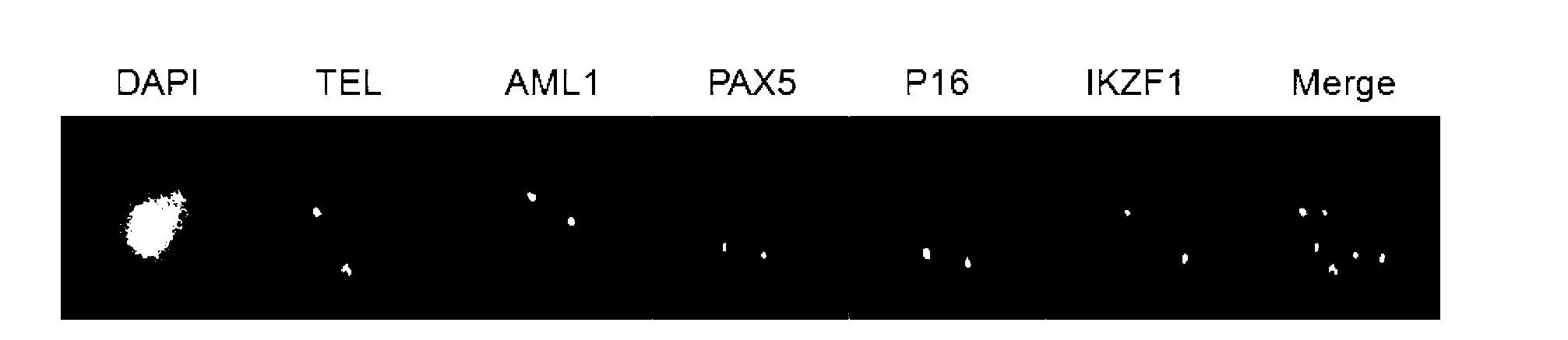
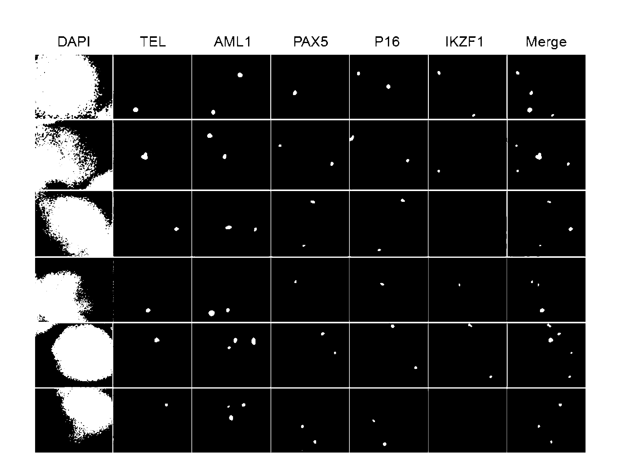
[0133] The results of fluorescence in situ hybridization showed that in the same nucleus, each probe obtained two clear fluorescent signals, and 5 probes obtained a total of 10 corresponding fluorescent signal points. Green-TEL (green), PF555-AML1 (red), PF590-PAX5 (orange), HyPer5-P16 (purple), PF415-IKZF1 (blue) multicolor images obtained from ( image 3 ), it can be seen that the fluorescence signals of the five different genes detected at the same time are all strong, the images are clear, and the signal-to-noise ratio is high. The method is reliable and stable. Embodiment 3: A fluorescent in situ hybridization detection kit for acute lymphoblastic leukemia (50 parts), the composition is as follows:
[0134] 1) Fluorescence-labeled probe set hybridization mixture 100 microliters; 1 tube
[0135] 2) DAPI counterstain solution 500 microliters; 1 tube
[0136] 3) 20ml of 20X SSPE washing solution; 1 bottle
[0137] 4) 1 instruction manual.
[0138] The concentration of t...
PUM
 Abstract
Description
Claims
Application Information