Fiber-based non-woven biodegradable ureteral stent tube and preparation method thereof

A ureteral stent tube and a biodegradable technology are applied in the field of fiber-based non-woven biodegradable ureteral stent tube and its preparation field, which can solve problems such as ureteral obstruction, achieve good biocompatibility, good product identity, and high economic benefits Effect

Inactive Publication Date: 2014-04-16
SHANGHAI CHILDRENS HOSPITAL +1
View PDF8 Cites 0 Cited by
  • Summary
  • Abstract
  • Description
  • Claims
  • Application Information

AI Technical Summary

Problems solved by technology

[0009] The technical problem to be solved by the present invention is to solve the problem of ureteral obstruction caused by the degradation of existing degradable ureteral stents

Method used


Image

  • Fiber-based non-woven biodegradable ureteral stent tube and preparation method thereof
  • Fiber-based non-woven biodegradable ureteral stent tube and preparation method thereof
  • Fiber-based non-woven biodegradable ureteral stent tube and preparation method thereof

Examples


Embodiment 1

[0031] Preparation of fiber raw materials: PGA and PLGA (PGA:PLA=90:10) chip raw materials, prepared by melt spinning process PGA (30-100dtex / 8-24f) multifilament fiber and PLGA (30-100dtex / 8-24f) Multifilament fiber; spinning is carried out on a micro spinning machine, and the spinning temperature is 255°C.

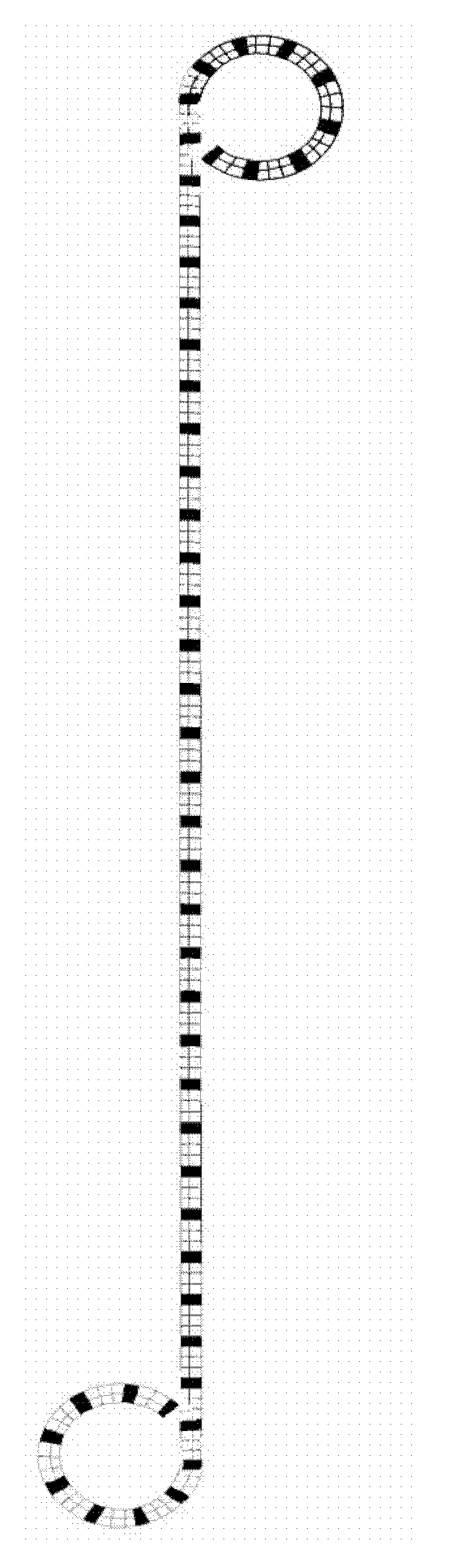
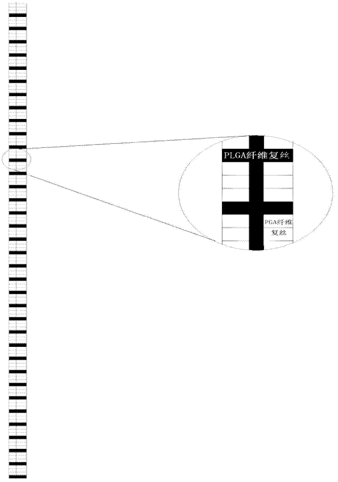
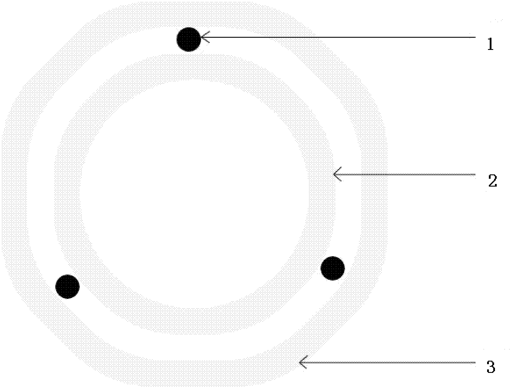
[0032] First arrange PLGA (30dtex / 8f) and PGA (30dtex / 8f) multifilament fibers in parallel in 1:3, as figure 2 , 3, wound in parallel on a mandrel with a diameter of 1.0mm, and then placed a PLGA fiber multifilament at intervals of 120° in the longitudinal axis direction, and wound a layer on its outer layer according to the above method. The total length is 16.0cm; set it in a vacuum oven at 210°C for 1min, and then remove the support tube layer from the mandrel; insert a 3.0cm-long tube with a diameter smaller than the inner diameter of the support tube and a length of 3.0cm at both ends of the preliminarily shaped support tube. The mandrel is wrapped around a polyt...

Embodiment 2

[0036] Preparation of fiber raw materials: PGA and PLGA (PGA:PLA=95:5) chip raw materials (wherein adding barium sulfate with a mass ratio of 10-20%), prepare PGA (30-100dtex / 8-24f) by melt spinning process And PLGA (30-100dtex / 8-24f) multifilament fibers; spinning is carried out on a micro-spinning machine, and the spinning temperature is 255°C. First arrange PLGA (30dtex / 8f) and PGA (30dtex / 8f) multifilament fibers in parallel at 1:5, such as figure 2 , 3 , wound in parallel on a mandrel with a diameter of 3.0mm, and then placed a PLGA fiber multifilament at intervals of 90° in the longitudinal axis direction, and wound a layer on its outer layer according to the above method. The total length is 44.0cm; set it in a vacuum oven at 210°C for 15 minutes, and then remove the stent tube layer from the mandrel; insert a 9.5cm long tube with a diameter smaller than the inner diameter of the stent tube at both ends of the preliminarily shaped stent tube. The mandrel is wrapped a...

Embodiment 3

[0040] Preparation of fiber raw materials: PGA and PLGA (PGA: PLA = 60: 40) chip raw materials (wherein adding barium sulfate with a mass ratio of 30%), prepare PGA (30-100dtex / 8-24f) and PLGA by melt spinning process (30-100dtex / 8-24f) multifilament fiber; the spinning is carried out on a micro-spinning machine made by Shanghai Tianqing Biological Materials Co., Ltd., and the spinning temperature is 255°C.

[0041] First arrange PLGA (30dtex / 8f) and PGA (30dtex / 8f) multifilament fibers in parallel in 1:5 on the braiding machine, such as figure 2 , 3 , wound in parallel on a mandrel with a diameter of 5.8mm, and then placed a PLGA fiber multifilament at intervals of 90° in the longitudinal axis direction, and wound a layer on its outer layer according to the above method. Then install the above method to co-wind 4 layers of fiber multifilament. The total length is 65.0cm; shape it in a vacuum oven at 210°C for 30 minutes, and then remove the stent tube layer from the mandre...


PUM

PropertyMeasurementUnit
thicknessaaaaaaaaaa
lengthaaaaaaaaaa
diameteraaaaaaaaaa

Abstract

The invention belongs to the technical field of bioengineering and particularly relates to a fiber-based non-woven biodegradable ureteral stent tube and a preparation method thereof. The fiber-based non-woven biodegradable ureteral stent tube disclosed by the invention is characterized by being in a 3D (Three Dimensional) latticed structure, wherein the 3D latticed structure consists of 30-100dtex / 8-24f PGA (polyglycolic acid) multifilament fiber and 30-100dtex / 8-24f PLGA (poly(lactic-glycolic acid)) multifilament fiber. The fiber-based non-woven biodegradable ureteral stent tube and the preparation method thereof have the advantages of simple preparation technology, higher economic benefits and good product identity; the biocompatibility and the mechanical performance of the stent tube are good; the degradation process is in step-by-step degradation according to time variation; and a degradation product is less than 0.1mm<3> and can be spontaneously excreted.

Description

technical field [0001] The invention belongs to the technical field of bioengineering, and in particular relates to a fiber-based non-woven biodegradable ureteral stent and a preparation method thereof. Background technique [0002] The main components of currently used ureteral stents are non-absorbable polyurethane, silicone rubber and metal materials, which need to be taken out again after surgery, and have poor tissue compatibility. Easy to form stones, infection, bleeding and tissue damage. If the stent tube is placed for a long time, it will cause serious consequences, such as loss of renal function or even nephrectomy. And a second operation is required to remove the indwelling stent. Cause multiple physical, mental and economical damages to patients, especially children patients who need to be pulled out after general anesthesia. [0003] The design of ureteral stents focuses on raw materials, surface characteristics, shape design, etc. An ideal ureteral stent sho...

Claims


Application Information

Patent Timeline
Patent Type & Authority Patents(China)
IPC IPC(8): A61L29/06A61L29/14B29C53/14
Inventor 陈方王璐尚亚峰王文祖周君梅王富军
Owner SHANGHAI CHILDRENS HOSPITAL
Why Eureka
Try Eureka