Access to the left atrium and reduction of mitral valve leaflet mobility

a technology of left atrium and mitral valve, which is applied in the field of cardiology and cardiac surgery, can solve the problems of congestive heart failure, low cardiac output, shortness of breath, etc., and achieve the effects of reducing the mobility of one, and quick, simple and accura

Inactive Publication Date: 2010-11-25
MOR RES APPL LTD
View PDF8 Cites 64 Cited by
  • Summary
  • Abstract
  • Description
  • Claims
  • Application Information

AI Technical Summary

Benefits of technology

[0029]The present invention, in some embodiments thereof, relates to the fields of cardiac surgery and cardiology. Some embodiments of the present invention relate to access of the left atrium or the mitral valve of a mammalian heart through the coronary sinus. Some embodiments of the present inventio

Problems solved by technology

Mitral valve insufficiency leads to many complications including arrhythmia, atrial fibrillation, cardiac palpitations, chest pain, congestive heart failure, fainting, fatigue, low cardiac output, orthopnea, paroxysmal nocturnal dyspnea, pulmonary edema, shortness of breath, and sudden death.
Initially, ischemic mitral regurgitation is a minor problem, typically leading only to shortness of breath during physical exercise due to the fact that a small fraction of b

Method used


Image

  • Access to the left atrium and reduction of mitral valve leaflet mobility
  • Access to the left atrium and reduction of mitral valve leaflet mobility
  • Access to the left atrium and reduction of mitral valve leaflet mobility

Examples


first embodiment

[0181]Reference is now additionally made to FIG. 5G, which is a schematic depiction of an optional configuration of the present invention relating to the minimally invasive conduit to the left atrium.

[0182]FIG. 5G depicts the elongated catheter body 72, the distal end 76, and the distal tip 78, depicts an angle 205 between the elongated catheter body 72 and the distal end 76. The angle 205 is used to negotiate the entrance to the coronary sinus, which is at an angle from the direction from which the elongated catheter body 72 typically enters the right atrium (12 of FIG. 1). The angle is optionally between about 70° and 110°.

[0183]While balloon 90 is in a first non-anchoring state, device 70 is mounted onto catheter-guiding guide wire 84 through catheter-guiding guide wire lumen 88 and directed in the usual way so that distal end 76 of elongated catheter body 72 is located inside coronary sinus 52 where side port 82 faces wall 98 of cardiac tissue separating coronary sinus 52 from l...

third embodiment

[0246]Reference is now additionally made to FIG. 9, which is a schematic depiction of the present invention relating to mitral valve leaflet augmentation.

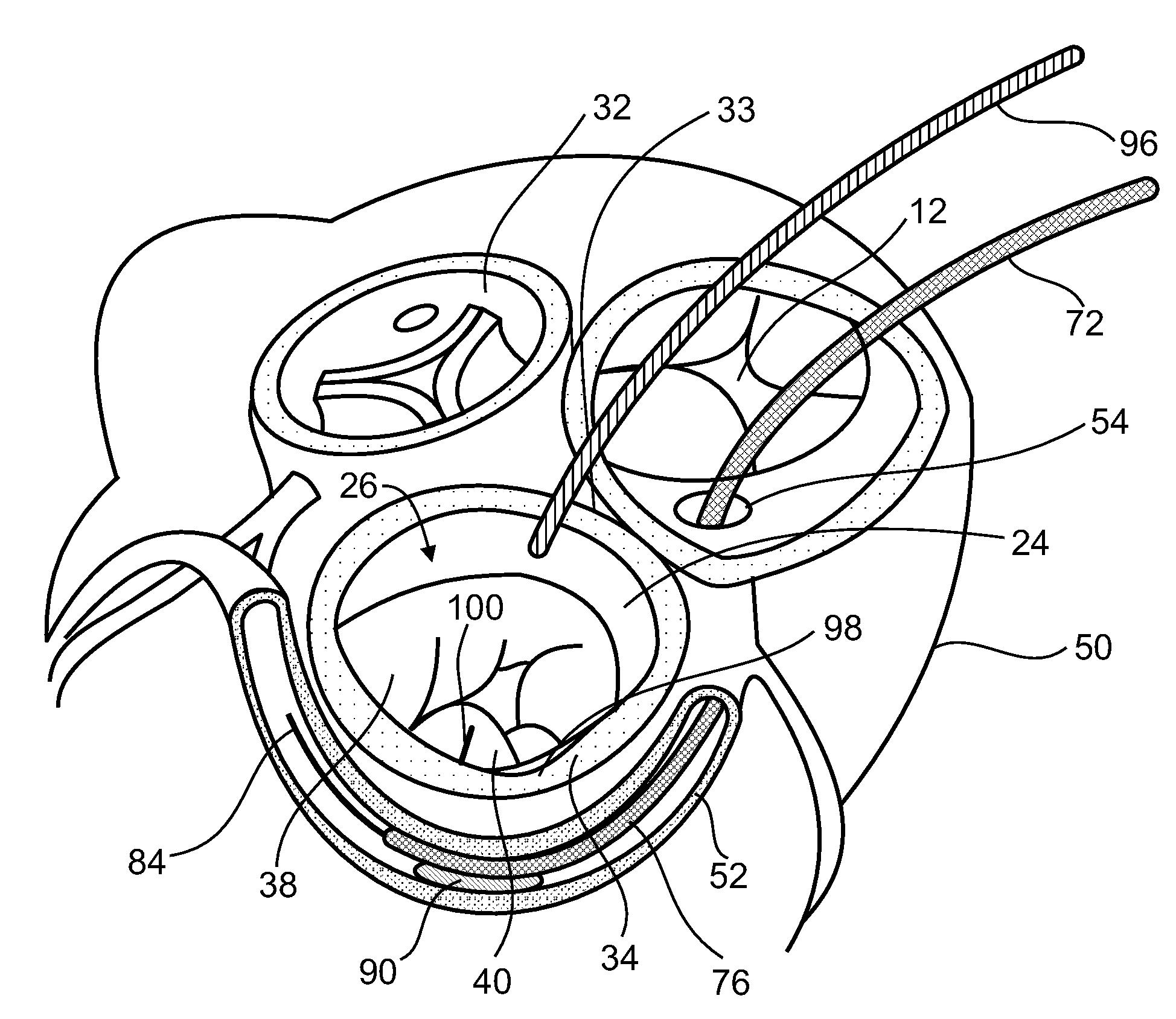
[0247]As described above with reference to other embodiments, an elongated catheter body 72 enters the left atrium 24 through the coronary sinus 52. In the embodiment of FIG. 9, the elongated catheter body 72 is used to deploy an obstructor deployment catheter 268. The obstructor deployment catheter 268 deploys an expandable obstructor 270, in the anterior leaflet 38.

[0248]In the embodiment discussed above, guidance of leaflet-engaging components 104 and 106 is performed with the help of optical observation through optical fiber bundle 96 that passes through the body of the subject including past the femoral vein, the inferior vena cava, right atrium 12 and into left atrium 24 through a transseptal puncture. In some embodiments, an optical fiber or similar component is positioned to guide leaflet-engaging components through a diffe...

fourth embodiment

[0267]Reference is now additionally made to FIG. 10A, which is a schematic depiction of the present invention relating to a mitral valve obstruction device.

[0268]The mitral valve anti-regurgitation device 220 includes an obstruction 221 connected to an extension wire 222, which is further connected to an anchor 223.

[0269]The anti-regurgitation device 220 passes through a catheter 225, at the end of a wire 224.

[0270]Reference is now additionally made to FIG. 10B, which is a schematic depiction of the fourth embodiment of FIG. 10A, deployed in a heart.

[0271]The anchor 223 is depicted as having been attached to a wall of the left ventricle 28. By way of a non-limiting example, the anchor 223 is optionally a sharp wire with a shape of a corkscrew, suitable for anchoring in the wall of the left ventricle. By way of another non-limiting example, the anchor 223 is optionally a staple, suitable for anchoring in the wall of the left ventricle.

[0272]The obstruction 221 of the anti-regurgitati...


PUM


Abstract

Disclosed are a method for engaging cardiac valve leaflets, including a) directing a distal end of an elongated catheter body of a leaflet-engaging device into a coronary sinus of a heart, b) passing a first leaflet-engaging component located proximate to the distal end of the elongated catheter body of the leaflet-engaging device through cardiac tissue separating the coronary sinus and a left atrium of the heart to enter a left atrium of the heart, and c) engaging a first cardiac valve leaflet with the first leaflet-engaging component, thereby engaging at least one cardiac valve leaflet. Related apparatus and methods are also described.

Description

RELATED APPLICATION / S[0001]This application claims priority from U.S. Provisional Patent Application No. 60 / 991,748, filed by the same Inventor on 2 Dec. 2007, and from US Provisional Patent Application No. 61 / 041,942 filed on 3 Apr. 2008.[0002]The contents of all of the above documents are incorporated by reference as if fully set forth herein.FIELD AND BACKGROUND OF THE INVENTION[0003]The present invention, in some embodiments thereof, relates to the fields of cardiac surgery and cardiology. Some embodiments relate to accessing the left atrium or the mitral valve of a mammalian heart through the coronary sinus. Some embodiments relate to the reduction of mobility of one or both mitral valve leaflets, for example to the extent that performance of surgical intervention to the mitral valve, for example, deployment of a leaflet-edge capturing device to perform a “bow-tie” repair of the mitral valve, is relatively quick, simple, and accurate.[0004]The human heart 10, depicted in cross-...

Claims


Application Information

Patent Timeline
IPC IPC(8): A61F2/24
CPCA61F2/2454A61F2/246A61F2/2463A61F2/2466A61B17/3478A61B2017/308A61B2017/00783A61B17/3421A61B2017/00243A61B2017/00247A61B2017/22098A61B2017/22069
Inventor ORLOV, BORIS
Owner MOR RES APPL LTD
Why Eureka
Try Eureka