Magnetic resonance angiography method and magnetic resonance angiography system

A vascular imaging and magnetic resonance technology, applied in the field of medical imaging, can solve the problems of hand and foot arterial difficulties, the influence of blood flow speed and direction, and display tortuousness, etc., to improve clinical practicability, improve effect, and improve imaging. fast effect

Inactive Publication Date: 2013-05-22
SHENZHEN INST OF ADVANCED TECH
View PDF2 Cites 18 Cited by
  • Summary
  • Abstract
  • Description
  • Claims
  • Application Information

AI Technical Summary

Problems solved by technology

However, the existing methods are very susceptible to the influence of blood flow velocity and direction, and it is very difficult to display the tortuous and small-diameter hand and foot arteries

Method used


Image

  • Magnetic resonance angiography method and magnetic resonance angiography system
  • Magnetic resonance angiography method and magnetic resonance angiography system
  • Magnetic resonance angiography method and magnetic resonance angiography system

Examples


Embodiment approach

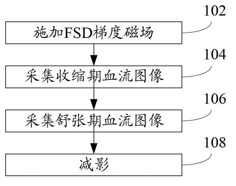
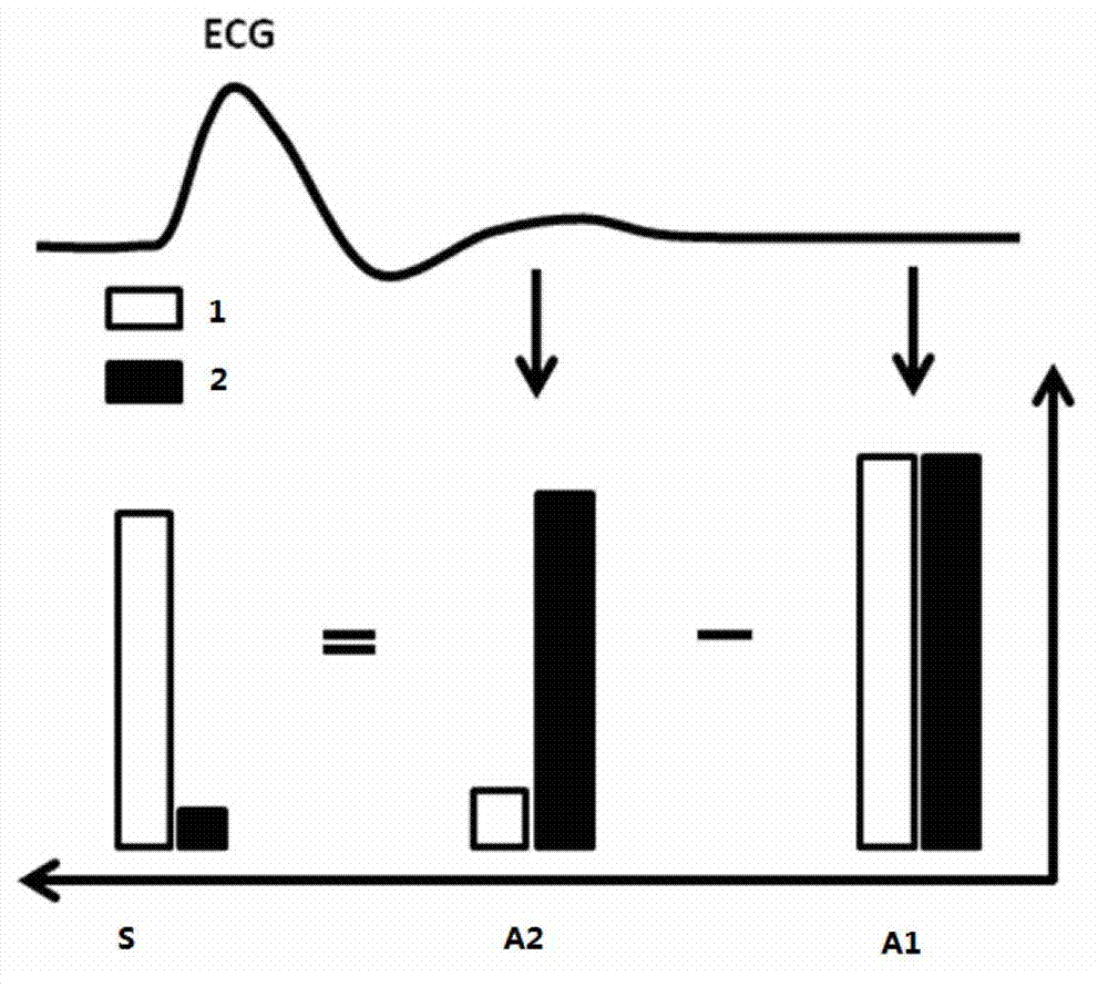
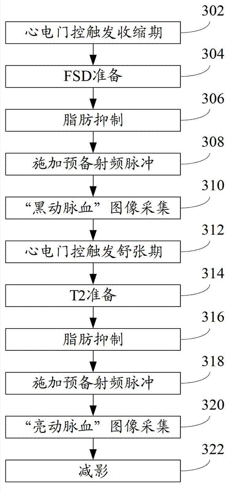
[0050] In one embodiment, step 102 specifically includes: during systole triggered by ECG gating, applying a 90° x -180° y -90° -x RF pulse sequence and symmetrical loading at 180° y Flow-sensitive phase gradient magnetic fields on both sides of the pulse, and a gradient magnetic field for removing residual magnetic moments is applied after the pulse sequence.

[0051] One embodiment, the first-order moment m of the flow-sensitive bulk gradient magnetic field 1 Satisfy minimum, where φ is the total spin phase, γ is the magnetic spin ratio constant, is the flow velocity of the ith spin.

[0052] An implementation manner, step 104 specifically includes:

[0053] Step A01: Carry out fat suppression using spectrally selective fat saturation technology;

[0054] Step A02: using 10 preparatory radio frequency pulses with linearly increasing flip angles;

[0055] Step A03: Using the equilibrium steady-state free precession technique to collect blood flow images during vasoc...


PUM


Abstract

The invention discloses a magnetic resonance angiography method. The magnetic resonance angiography method includes applying a flowing sensitive dispersed phase gradient magnetic field in the readout direction and / or the phase encoding direction and / or the layer selection direction in a vasoconstriction period and then applying a residual magnetic moment removing gradient magnetic field after the flowing sensitive dispersed phase gradient magnetic field is applied; collecting blood flow images in the vasoconstriction period; collecting blood flow images in a vasodilatation period; and carrying out subtraction on the blood flow images in the vasoconstriction period and the blood flow images in the vasodilatation period to obtain blood flow images. The invention further discloses a magnetic resonance angiography system. According to the magnetic resonance angiography method and the magnetic resonance angiography system, in the specific implementation method, the flowing sensitive dispersed phase is used in the vasoconstriction period imaging process, so that intra-arterial self-spin dispersed phase can significantly lower arterial blood signals, and therefore the influence on imaging due to the velocity and the direction of blood flow can be reduced, high-quality peripheral arterial images can be obtained, and clinical practicability of the non-enhanced magnetic resonance angiography technology is improved.

Description

technical field [0001] The present application relates to medical imaging, in particular to a magnetic resonance angiography method and system. Background technique [0002] Magnetic resonance angiography (Magnetic Resonance Angiography, MRA) is currently one of the main means of clinical diagnosis of vascular lesions, it usually includes enhanced MRA and non-enhanced MRA two methods. [0003] Enhanced MRA refers to the method of imaging blood vessels after intravascular injection of contrast agent (paramagnetic contrast agent containing gadolinium). In the past, it was believed that the side effects of gadolinium-containing paramagnetic contrast agents were very small and did not pose a threat to the human body. However, a large number of recent research data show that this type of contrast agent has potential damage to renal function, especially in patients with renal insufficiency, which can cause a fatal complication called nephrogenic systemic fibrosis. In addition, e...

Claims


Application Information

Patent Timeline
Patent Type & Authority Applications(China)
IPC IPC(8): A61B5/055
Inventor 刘新樊昭阳张娜钟耀祖郑海荣李德彪
Owner SHENZHEN INST OF ADVANCED TECH
Why Eureka
Try Eureka