Intravascular ultrasound image segmentation method

A technology of ultrasonic image and image segmentation, applied in the field of medical image processing, can solve problems such as fast correction, inconvenient results, complex modeling process, etc., and achieve the effect of ensuring automation

Inactive Publication Date: 2013-06-19
UNIV OF SHANGHAI FOR SCI & TECH +1
View PDF4 Cites 18 Cited by
  • Summary
  • Abstract
  • Description
  • Claims
  • Application Information

AI Technical Summary

Problems solved by technology

At present, there are three main categories of computer automatic segmentation algorithms for intravascular ultrasound images: the first category is statistical methods (G. Mendizabal-Ruiz, M. Rivera, et al., “A probabilistic segmentation method for the identification of luminal borders in intravascular ultrasound images”, IEEE Conference on Computer Vision and Pattern Recognition, pp.1-8, 2008.), statistical modeling of the gray distribution of images to achieve intravascular ultrasound image segmentation, but artifacts in intravascular ultrasound images Complex image features such as calcification and calcification will greatly reduce the accuracy of statistical modeling; the second category achieves intravascular ultrasound image segmentation by means of machine learning (1.E. G. Bovenkamp, ​​J. Dijkstra, J. G. Bosch, et al., “Multi -agent segmentation of IVUS images”, Patten Recognition, Vol.37, No.4, pp.647-663, 2004; 2. G. Unal, S. Bucher, S. Carlier, et al., “Shape-driven segmentation of the arterial wall in intravascular ultrasound images", IEEE Trans. On information technology in biomedicine, Vol.12, No.3, pp.335-346, 2008.), the model of this type of method is complicated, and there are many limitations in practical application; The third category is the method based on the active contour model (1. Zhang Qi, Wang Yuanyuan, etc., "Active Contour Model and Contourlet Multi-resolution Analysis to Segment Intravascular Ultrasound Images", Optical Precision Engineering, Vol.16, No.11, pp.2301-311, 2008; 2. X. Zhu, P. Zhang, J. Shao, et al., “A snake-based method for segmentation of intravascular ultrasound images and its in vivo validation”, Ultrasonics, Vol .51, pp.181-189, 2011.), this kind of square often needs to give the initial contour line, and the segmentation result is easily affected by complex image features such as noise and different patches
Although the above-mentioned types of intravascular ultrasound image segmentation methods have a high degree of automation, they often need to go through a very complicated modeling process, and it is not convenient to quickly correct the results through human-computer interaction.

Method used


Image

  • Intravascular ultrasound image segmentation method
  • Intravascular ultrasound image segmentation method
  • Intravascular ultrasound image segmentation method

Examples


Embodiment Construction

[0037] The present invention will be described in further detail below in conjunction with the accompanying drawings.

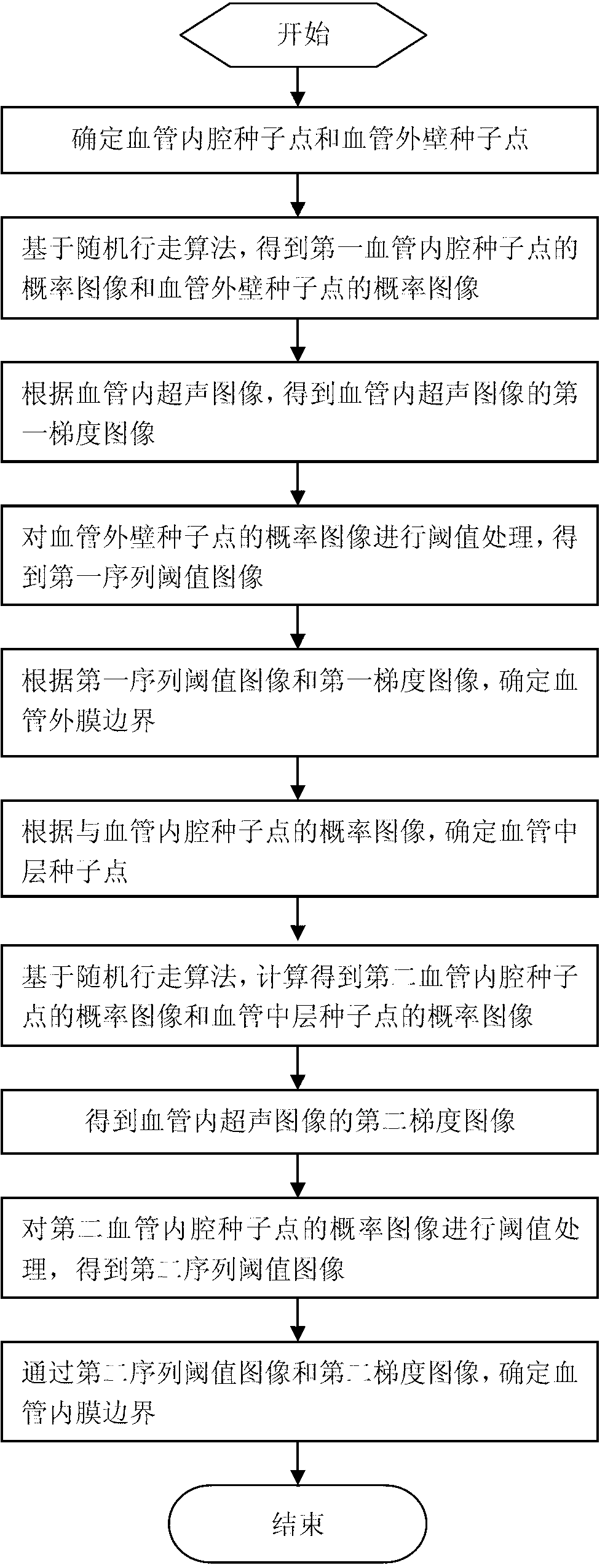
[0038] figure 1 It is a flow chart of the intravascular ultrasound image segmentation method of the present invention.

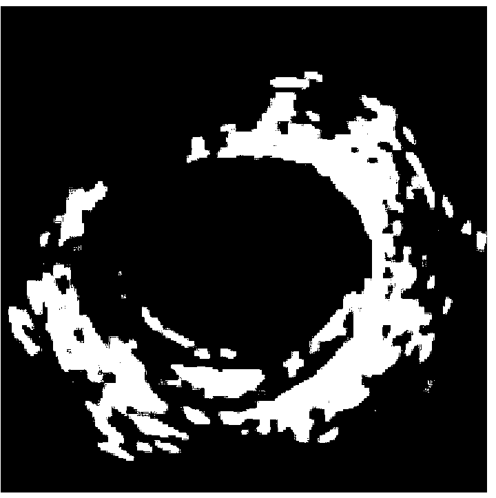
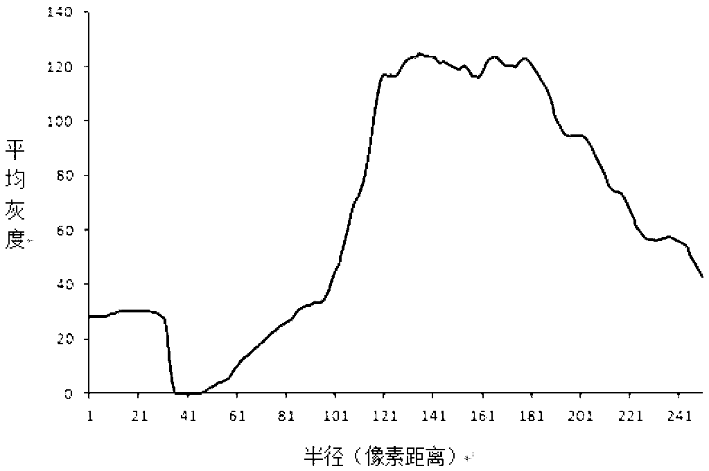
[0039] figure 2 is the intravascular ultrasound image of this embodiment, image 3 is the average grayscale curve of the intravascular ultrasound image in this embodiment. like image 3 As shown, the average grayscale curve of the intravascular ultrasound image takes the center point of the intravascular ultrasound image as the zero point coordinate point, the radius of each circle with the center point as the center as the abscissa, and all pixels on each circle The average gray value of the point is the ordinate. Figure 4 It is a schematic diagram of the seed point of the lumen of the blood vessel and the seed point of the outer wall of the blood vessel in this embodiment. like Figure 4 As shown, the average grayscale curve of t...


PUM


Abstract

An intravascular ultrasound image segmentation method includes the steps: determining seed points of blood vessel lumens and seed points of the outer wall of a blood vessel; obtaining probability images of the seed points of a first blood vessel lumen and the seed points of the outer wall of the blood vessel; obtaining a first gradient image of a intravascular ultrasound image; processing a threshold of the probability image of the seed points of the outer wall of the blood vessel to obtain a first sequence threshold image; determining an outer membrane boundary of the blood vessel according to the first sequence threshold image and the first gradient image; taking second communication area boundary pixels as seed points of a media; obtaining probability images of the seed points of a second blood vessel lumen and the seed points of the media; obtaining a second gradient image of the intravascular ultrasound image; processing a threshold of the probability image of the seed points of the second blood vessel lumen to obtain a second sequence threshold image; and determining an inner membrane boundary of the blood vessel according to the second sequence threshold image and the second gradient image. The probability of the probability images of the seed points of the blood vessel lumens in a second communication area is higher than 0.5.

Description

technical field [0001] The present invention relates to the field of medical image processing, in particular to a random walk (Random Walker) algorithm-based method applied to intravascular ultrasound (IVUS: Intravascular ultrasound) image segmentation. Background technique [0002] Intravascular Ultrasound (IVUS: Intravascular Ultrasound), as an interventional real-time ultrasound imaging technology, can not only display the shape of the vascular lumen, but also display the layered structure of the vascular wall. very important value. The diagnosis of atherosclerosis based on IVUS requires the acquisition of quantitative indicators of image features of atherosclerosis, such as vascular lumen area and plaque area. The accurate extraction of these quantitative indicators depends on effective image segmentation. Manual segmentation means that doctors manually delineate the lumen of blood vessels, the boundaries of media and adventitia, etc., which is not only time-consuming a...

Claims


Application Information

Patent Timeline
Patent Type & Authority Applications(China)
IPC IPC(8): G06T7/00
Inventor 严加勇崔崤峣
Owner UNIV OF SHANGHAI FOR SCI & TECH
Why Eureka
Try Eureka