Preparation method of microchip for microprotein detection

A micro-protein and microchip technology, applied in the fields of plasma separation and protein detection, can solve problems such as reduced separation efficiency, blockage of filter membrane pores, and sample contamination.

Active Publication Date: 2013-09-04
南京艾拓生命科技有限公司
View PDF7 Cites 5 Cited by
  • Summary
  • Abstract
  • Description
  • Claims
  • Application Information

AI Technical Summary

Problems solved by technology

However, these two methods have certain shortcomings. The former separation equipment is bulky and complicated to operate, and the latter filter membrane pores are easily blocked by blood cells, resulting in reduced separation efficiency and sample contamination.

Method used


Image

  • Preparation method of microchip for microprotein detection
  • Preparation method of microchip for microprotein detection
  • Preparation method of microchip for microprotein detection

Examples


Embodiment 1

[0029] Plasma Separation and Protein Detection of Healthy Whole Blood

[0030] Using a microfluidic system to separate plasma from whole blood of healthy people and detect proteins, the specific steps are as follows:

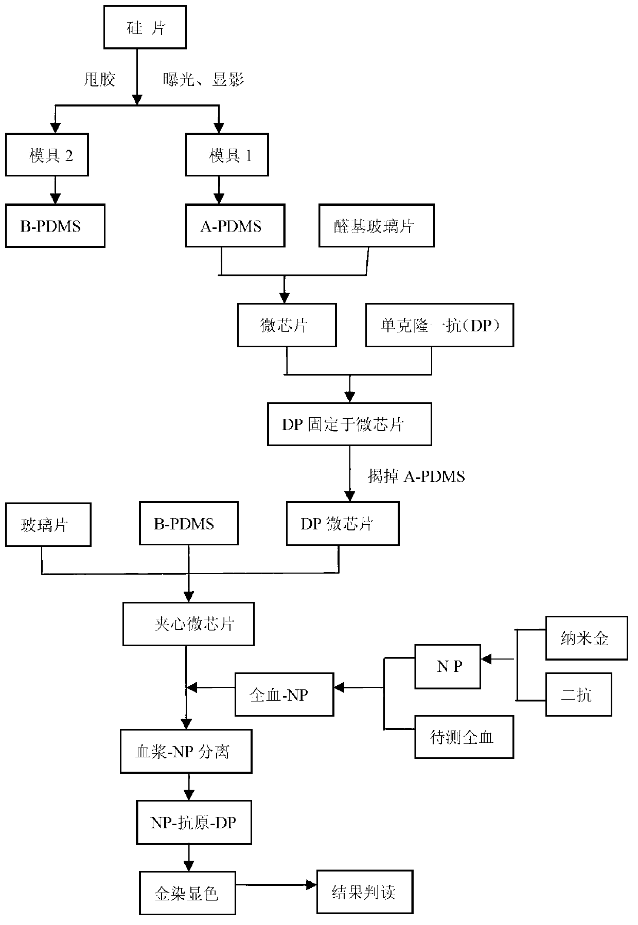
[0031] (1) Microfluidic chip preparation and protein immobilization

[0032] 1. Silicon wafer is used as the base material, and SU-8 photoresist is used as the mask layer to expose and develop the mold A for immobilizing the protein and the mold B for separation and detection;

[0033] 2. Mix PDMS and curing agent (Sylgard184curing agent) at a ratio of 10:1 (weight ratio), pour on mold A and mold B after removing air bubbles, heat and cure at 90°C for 1 hour, and make a microstructure;

[0034] 3. Peel off the PDMS block (A-PDMS) on mold A, punch the sample inlet and outlet holes with a punching needle, and then reversibly bond them together with the aldehyde-modified glass sheet;

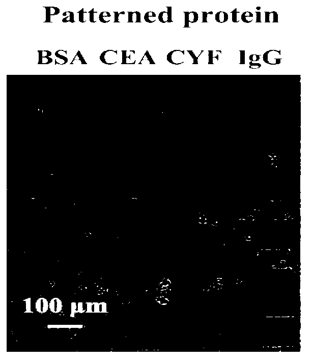
[0035] 4. Add 4 different primary antibody solutions (BSA, CEA, CyFRA21-1 and ...

Embodiment 2

[0050] Clinical whole blood plasma separation and protein detection

[0051] (1) Preparation of microfluidic chip

[0052] Same as (1) in Example 1.

[0053] (2) Preparation of gold nanoparticles and antibody labeling

[0054] Same as (2) in Example 1.

[0055] (3) Testing and interpretation of results

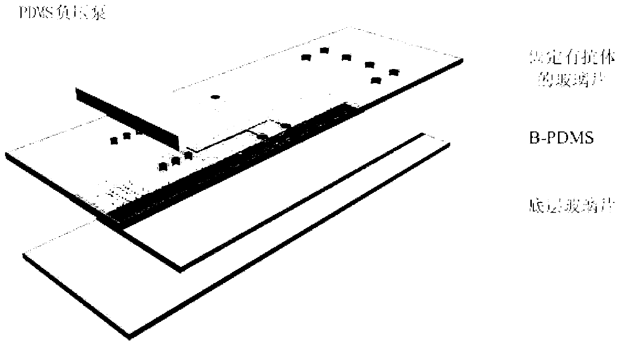
[0056] 1. Put the negative-pressured PDMS block in the sample hole of the sandwich microchip prepared above.

[0057] 2. Mix 10 μl of fresh lung cancer whole blood (contains CEA and CyFRA21-1 antigens identified in it), 0.5 μl of NP probe and mix evenly, take 3 μl of whole blood-NP mixed solution and add it to the injection port of the chip, under the action of negative pressure The blood flows into the microchannel, the blood cells naturally settle in the channel, the plasma / NP mixture flows into the detection area after separation, and forms a sandwich structure with the protein immobilized on the surface of the chip (i.e. NP-antigen-protein microchip), incubate at 37°C ...


PUM

PropertyMeasurementUnit
sizeaaaaaaaaaa

Abstract

The invention relates to a preparation method of a microchip for microprotein detection. The preparation method comprises the following steps of: respectively exposing and developing a silicon wafer taken as a substrate material and SU-8 photoresist taken as a masking layer so as to manufacture a mold A and a mold B; mixing PDMS (Polydimethylsiloxane) with a curing agent, respectively pouring the mixture into a mold A and a mold B, heating and curing; respectively stripping A-PDMS and B-PDMS; punching the A-PDMS, gluing a processed glass sheet to the punched A-PDMS; and stripping away the A-PDMS, aligning and gluing the glass sheet on which an antibody is fixed with the B-PDMS, and gluing another punched glass sheet on the other surface of the B-PDMS, thus obtaining the microchip. The microchip can be used for integrating the separation and the detection of blood plasma in whole blood and detecting multiple targets, has the characteristics of specificity, rapidness and high sensitivity and can be expected to be applied to the clinical diagnosis and detection of multiple proteins in trace amounts of whole blood.

Description

technical field [0001] The invention belongs to the field of blood plasma separation and protein detection, and in particular relates to a preparation method of a microchip for detection of trace protein. Background technique [0002] Whole blood is composed of liquid plasma, red blood cells, white blood cells, platelets and other components. In modern medical testing, due to the great interference of blood cells or hemoglobin on spectral analysis, plasma is usually separated from blood samples first, and then used for biochemical or immunodiagnostic analysis. At present, centrifugation and filtration are the most common methods used in clinical practice. The centrifugation method uses the principle of inertial centrifugal force and material sedimentation coefficient to achieve the separation effect, while the filtration method realizes the separation of plasma through a filter membrane or a solid support. However, these two methods have certain shortcomings. The former se...

Claims


Application Information

Patent Timeline
Patent Type & Authority Applications(China)
IPC IPC(8): G01N33/68
Inventor 张宏莲李刚廖玲英毛红菊金庆辉赵建龙
Owner 南京艾拓生命科技有限公司
Why Eureka
Try Eureka