High-throughput immunohistochemical detection method and multi-sample immunohistochemical detection board

An immunohistochemistry, multi-sample technology, applied in the field of immunization, can solve the problems of time-consuming, labor-intensive, and high cost, achieve good application prospects, improve the effect of high-throughput immunohistochemical detection with a very low R&D success rate

Inactive Publication Date: 2013-10-09
成都安铂奥金生物科技有限公司
View PDF9 Cites 18 Cited by
  • Summary
  • Abstract
  • Description
  • Claims
  • Application Information

AI Technical Summary

Problems solved by technology

However, the existing immunohistochemical detection method is only suitable for the detection of one antibody, so as many candidate antibodies (hybridomas) as t

Method used


Image

  • High-throughput immunohistochemical detection method and multi-sample immunohistochemical detection board
  • High-throughput immunohistochemical detection method and multi-sample immunohistochemical detection board
  • High-throughput immunohistochemical detection method and multi-sample immunohistochemical detection board

Examples


Embodiment 1

[0053] Example 1: High-throughput immunohistochemical detection of surgical resection or biopsy tissue

[0054] (1) Preparation of tissue slices to be tested: Dot matrix tissue slices are prepared by conventional tissue microarray technology, and the diameter, spacing, and distribution position of each tissue completely match the porous matrix plate.

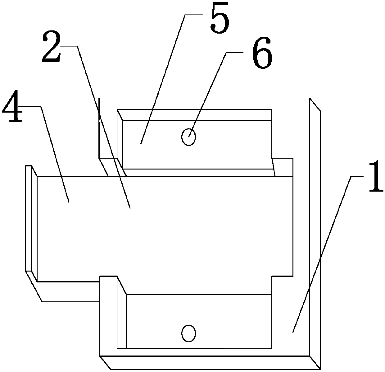
[0055] (2) Mount the glass slide: put the tissue chip slide treated with wax removal, hydration, antigen retrieval and non-specific antigen blocking into the slide fixing groove of the base, and confirm that the tissue section faces upward.

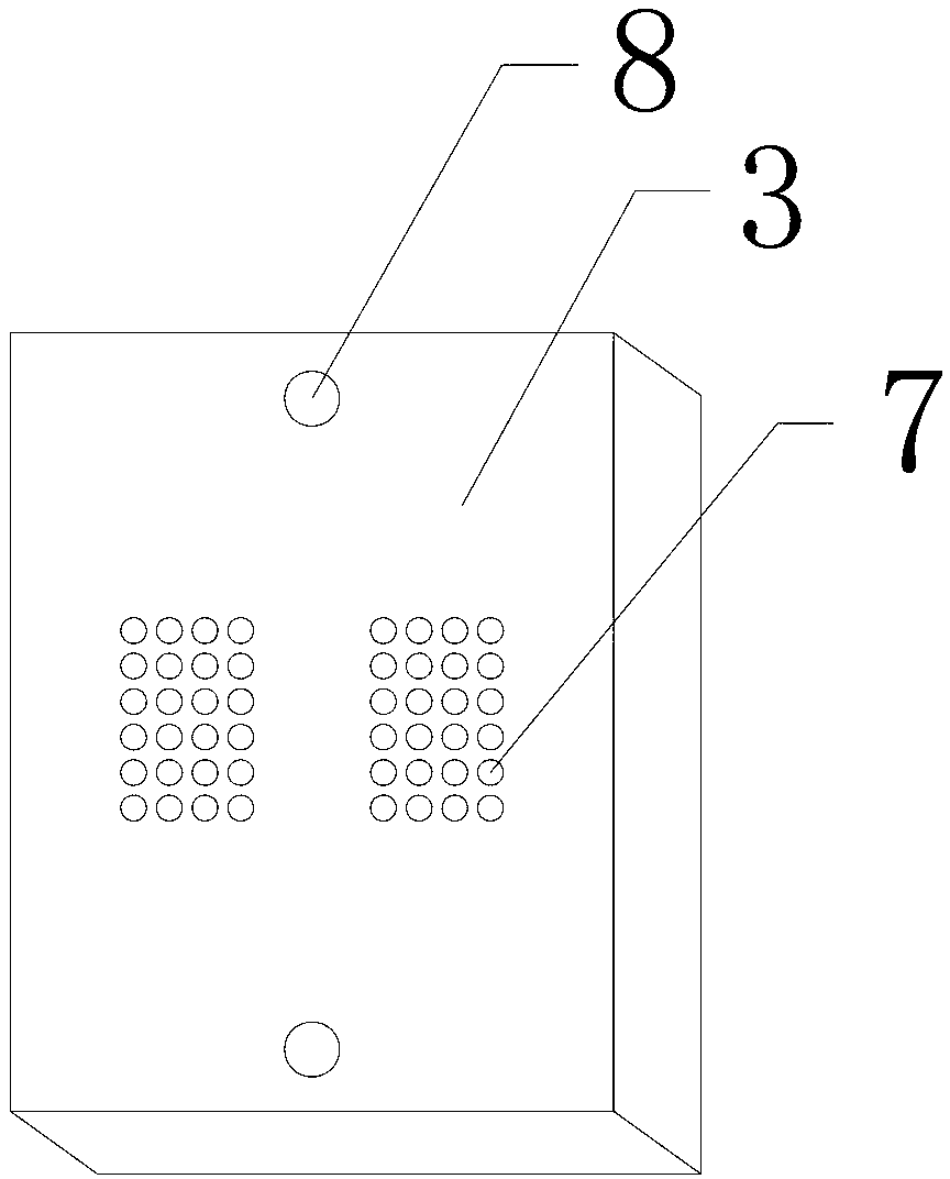
[0056] (3) Install the porous matrix plate: Put the waterproof pad of the porous matrix plate into the fixed groove of the porous plate with one side facing down, adjust each tissue section point to be completely inserted into the corresponding small hole of the porous matrix plate and fasten the bolt .

[0057] (4) Add the supernatant of the original hybridoma culture medium into the corr...

Embodiment 2

[0060] Example 2: Immunohistochemical detection of tissue chips made from primary cultured cell lines or transfected cells

[0061] (1) Preparation of paraffin-embedded cell tissue blocks:

[0062] 1) Harvest the culture, centrifuge and wash the cells twice with phosphate buffer;

[0063] 2) Suspend the cells in 4% formalin fixative, incubate at 4°C for 1-2 hours, then centrifuge and discard the supernatant;

[0064] 3) Suspend the cells in phosphate buffer and incubate at 37°C for 10 minutes;

[0065] 4) Mix the cell suspension with an equal amount of 3% agarose gel solution at 50°C;

[0066] 5) Immediately add the new cell and agarose gel suspension into the tissue block preparation mold, immediately centrifuge for 5 minutes, decompose the tissue block preparation mold, and take out the prepared cell line tissue block;

[0067] 6) The obtained cell tissue block was cut into 1.0 mm thick, and then dehydrated and paraffin-embedded.

[0068] (2) Preparation of conventional ...

experiment example 1

[0075] Experimental example 1: Screening of Napsin A immunohistochemical monoclonal antibody

[0076] Napsin A is an aspartic protease. Among the malignant tumors of the lung, more than 80% of lung adenocarcinoma patients showed Napsin A positive in biopsy pathological examination, while all lung squamous cell carcinoma and small cell lung cancer patients showed Napsin A negative in pathological examination. Therefore, Napsin A immunohistochemical monoclonal antibody has become an important reagent for the differential diagnosis of lung cancer and the diagnosis of lung adenocarcinoma metastasis. However, Napsin A monoclonal antibody for immunohistochemical diagnosis is extremely difficult to prepare. So far, there has not been a generally recognized Napsin A monoclonal antibody for immunohistochemistry in the international market.

[0077] Adopt the method of the present invention to screen Napsin A monoclonal antibody as follows:

[0078] 1. Experiment preparation

[0079...


PUM

PropertyMeasurementUnit
Horizontal sizeaaaaaaaaaa

Abstract

The invention discloses a multi-sample immunohistochemical detection board, and also discloses a high-throughput immunohistochemical detection method with the detection board. The method comprises the following steps of: (1) preparing a tissue section to be detected; (2) flatly putting the tissue section to be detected in a step (1) in a transverse groove (2) after dewaxing, hydrating, carrying out antigen retrieval, and carrying out closed treatment; (3) putting a perforated plate (3) in a longitudinal groove (5) to be fixed, wherein one surface covered with a flexible waterproof lining puts down; (4) adding various antibody solutions to be detected to different through holes (7) for incubation; (5) carrying out color development on the tissue section to be detected, sealing the tissue section, and observing. The invention also discloses a mold for preparing a cell tissue block. The high-throughput immunohistochemical detection method can simultaneously carry out the detection on various antibodies on a same slide and has a good application prospect.

Description

technical field [0001] The invention relates to a high-throughput immunohistochemical detection method and a multi-sample immunohistochemical detection board, belonging to the field of immunity. Background technique [0002] As we all know, pathological diagnosis is the gold standard for clinical disease diagnosis. The modern conventional pathological diagnosis method is realized by staining the pathological tissue sections fixed on the slide with specific monoclonal antibodies, that is, immunohistochemical technique (immunohistochemical staining technique). [0003] Conventional immunohistochemical technology is developed on the basis of classic pathological section technology, and usually a slide contains only one type of pathological tissue section. The recently developed tissue chip technology can upload a variety of pathological tissue slices onto a standard glass slide, which solves the problem that only one type of pathological tissue can be loaded on a slide, and ac...

Claims


Application Information

Patent Timeline
IPC IPC(8): G01N33/577G01N33/531
Inventor 陈柏华李平韩星
Owner 成都安铂奥金生物科技有限公司
Why Eureka
Try Eureka