Method for preparing induced pluripotent stem cells using microvesicles derived from embryonic stem cells

A technology of pluripotent stem cells and embryonic stem cells, applied in embryonic cells, animal cells, germ cells, etc., can solve the problems of damaged cells, difficult mRNA synthesis process, induction of RNA immune response, etc., to achieve low damage and high delivery. Efficiency, loss prevention effect

Inactive Publication Date: 2014-01-01
POSTECH ACAD IND FOUND
View PDF2 Cites 5 Cited by
  • Summary
  • Abstract
  • Description
  • Claims
  • Application Information

AI Technical Summary

Problems solved by technology

[0004] In addition, methods to examine cell fusion (cellular, cytoplasmic) have the following disadvantages: many cells die due to induction of fusion using polyethylene glycol (PEG), which can damage cells to help large-sized cells fuse
However, those skilled in the art have found that mRNA is prone to degradation and difficult to synthesize, induces an immune response to RNA, and delivery of protein alone cannot achieve complete dedifferentiation

Method used


Image

  • Method for preparing induced pluripotent stem cells using microvesicles derived from embryonic stem cells
  • Method for preparing induced pluripotent stem cells using microvesicles derived from embryonic stem cells
  • Method for preparing induced pluripotent stem cells using microvesicles derived from embryonic stem cells

Examples


Embodiment 1

[0045] Example 1. Preparation of Embryonic Stem Cell-Derived Microvesicles

[0046] Take 5×10 6 Resuspend mouse embryonic stem cells in 3 ml of phosphate-buffered saline (PBS) solution at a concentration of 1 cell / ml. The resuspension was passed through a membrane filter with a pore size of 10 μm 10 times and a membrane filter with a pore size of 5 μm 10 times. 1ml50% OptiPrep TM , 1ml5% OptiPrep TM and 3ml of the cell suspension that passed through the membrane filter were placed in a 5ml ultracentrifuge tube. Subsequently, ultracentrifugation was performed at 100,000×g for 2 hours. At 50% OptiPrep TM and 5% OptiPrep TM Microvesicles are obtained in the interlayer.

Embodiment 2

[0047] Example 2. Characterization of microvesicles derived from embryonic stem cells

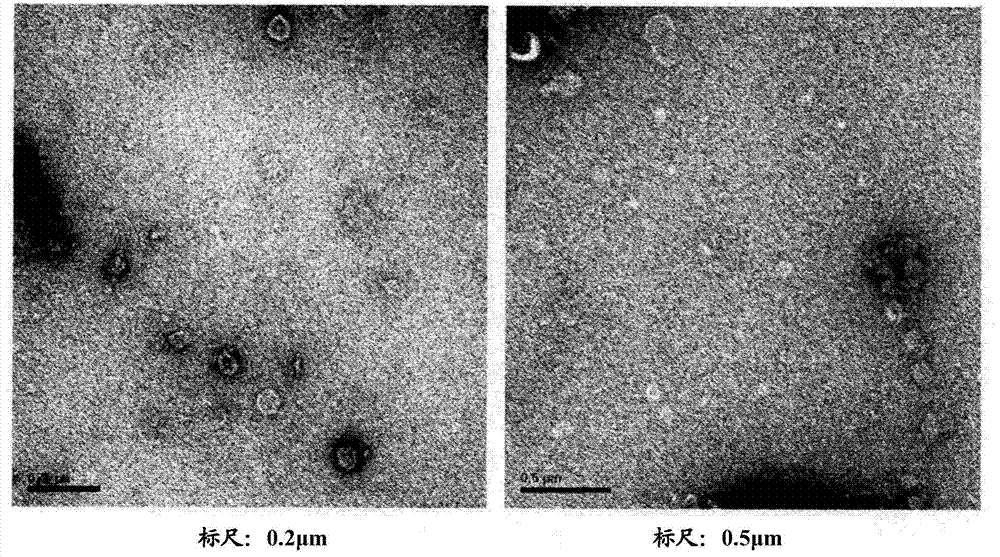
[0048] Microvesicles prepared from embryonic stem cells according to the method described in Example 1 were adsorbed on the glow-discharged carbon-coated copper grid for 3 minutes. The mesh was washed with distilled water and stained with 2% uranyl acetate for 1 minute, and the results observed using a transmission electron microscope JEM101 (Jeol, Japan) were as follows: figure 1 shown.

[0049] Such as figure 1 As shown in the TEM images of , it can be seen that microvesicles prepared from embryonic stem cells by extrusion are composed of lipid bilayers and generally form spherical shapes with a size of 100 nm to 200 nm.

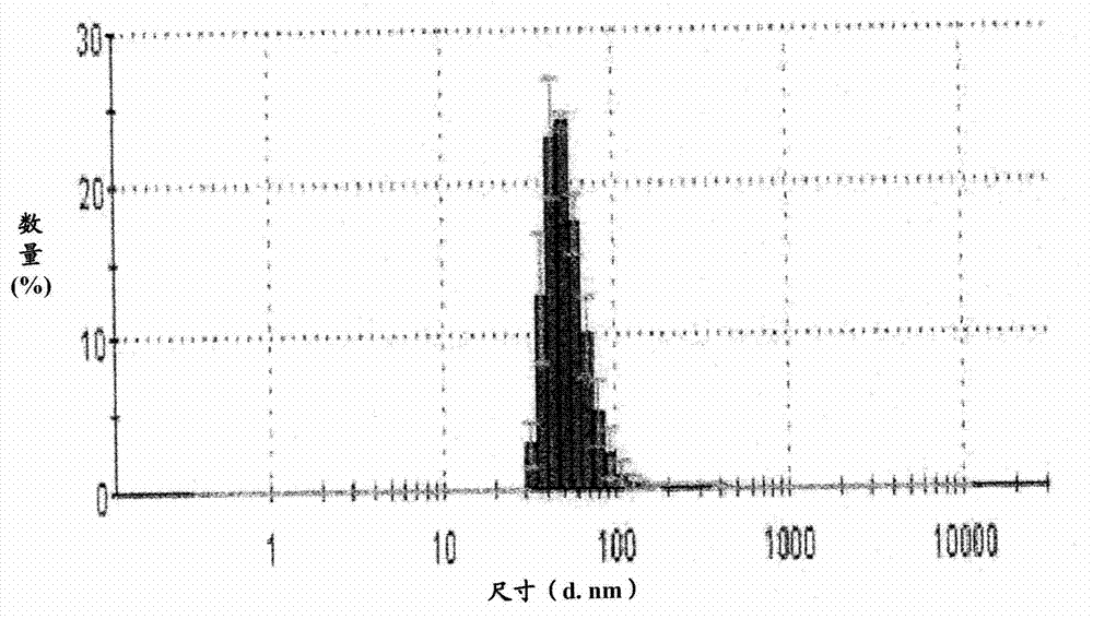
[0050] The microvesicles prepared from embryonic stem cells described in Example 1 were diluted in 1 ml of PBS to a concentration of 5 μg / ml. 1ml PBS containing microvesicles was placed in a cuvette and analyzed using a dynamic light scattering particle size analyz...

Embodiment 3

[0056] Example 3. Dedifferentiation of Somatic Cells Using Embryonic Stem Cell-Derived Microvesicles

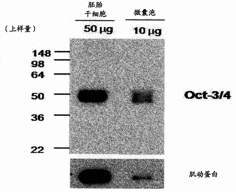
[0057] Coat a 6-well plate with 0.1% gelatin and inoculate 8×10 4 NIH3T3 cells, incubate the cells for 24 hours. Subsequently, each well was washed with PBS, and 2 ml of microvesicles prepared from embryonic stem cells according to the method described in Example 1 were diluted in fibroblast medium (DMEM, 10% FBS, 100 U / ml penicillin-streptomycin) , to a concentration of 100 μg / ml, which was then used to treat the incubated NIH3T3 cells. After 48 hours, approximately 2 to 3 cell colonies per well were recognized, and each cell colony was approximately 10 to 100 μm in size. Cell colonies were observed using an electron microscope, and the results were as follows Figure 5 shown.

[0058] Figure 5 The results shown demonstrate the dedifferentiation of NIH3T3 cells using embryonic stem cell-derived microvesicles to induce cell colonies. Wash each well with PBS and add 4...


PUM

PropertyMeasurementUnit
sizeaaaaaaaaaa

Abstract

The present invention relates to a method for dedifferentiating somatic cells using microvesicles derived from embryonic stem cells. More particularly, the present invention relates to a method for preparing pluripotent stem cells by treating somatic cells with a composition having microvesicles derived from embryonic stem cells. The method for preparing pluripotent stem cells according to the present invention effectively facilitates the dedifferentiation of somatic cells without side-effects using the microvesicles, and further, is expected to be significantly useful in the development of cell therapy products having immune-compatibility suitable for specific individuals.

Description

technical field [0001] The present invention relates to a method for producing induced pluripotent stem cells by dedifferentiating somatic cells using embryonic stem cell-derived microvesicles. Background technique [0002] Dedifferentiation is the process by which mature somatic cells revert to younger stem cells and dedifferentiation is involved in regeneration in vivo, which occurs in insects, amphibians, plants. Said dedifferentiation does not occur naturally in mammals such as humans, but only occurs artificially in mammals such as humans. Methods for dedifferentiation of cells start with methods using cell fusion. The first method to dedifferentiate somatic cells discovered in the art was to use a method characterized by the fact that embryonic stem cells (ESCs) become the dominant cells when they fuse with somatic cells. Since then, research has progressed in inducing dedifferentiation of somatic cells by fusing them with stem cells other than ESCs having abilities ...

Claims


Application Information

Patent Timeline
Patent Type & Authority Applications(China)
IPC IPC(8): C12N5/0735C12N5/10A61K35/54
CPCC12N5/0696C12N2502/02A61K35/545C12N2506/13A61P43/00
Inventor 郑多英金俊镐朴宰成李南雨高用柗金润根张寿哲崔银廷
Owner POSTECH ACAD IND FOUND
Why Eureka
Try Eureka