CT/FT/PET three-mode synchronous imaging device

A synchronous imaging, three-modality technology, used in diagnostic recording/measurement, medical science, sensors, etc., can solve the problems of shallow imaging depth, inability to detect early tumors, poor imaging effect, etc., and achieve accurate structural and functional information. Effect

Inactive Publication Date: 2014-05-28
XIDIAN UNIV
View PDF7 Cites 10 Cited by
  • Summary
  • Abstract
  • Description
  • Claims
  • Application Information

AI Technical Summary

Problems solved by technology

However, the imaging depth of FT is relatively shallow, and generally only 2-3cm imaging targets can be distinguished; PET can distinguish deep targets well, but the imaging effect is

Method used


Image

  • CT/FT/PET three-mode synchronous imaging device
  • CT/FT/PET three-mode synchronous imaging device
  • CT/FT/PET three-mode synchronous imaging device

Examples


Embodiment Construction

[0027] The present invention will be further described below in conjunction with the accompanying drawings. It should be noted that this embodiment is implemented on the premise of the technical solution of the present invention, and the detailed implementation and specific operation process are given, but the protection scope of the present invention is not limited to the following embodiments.

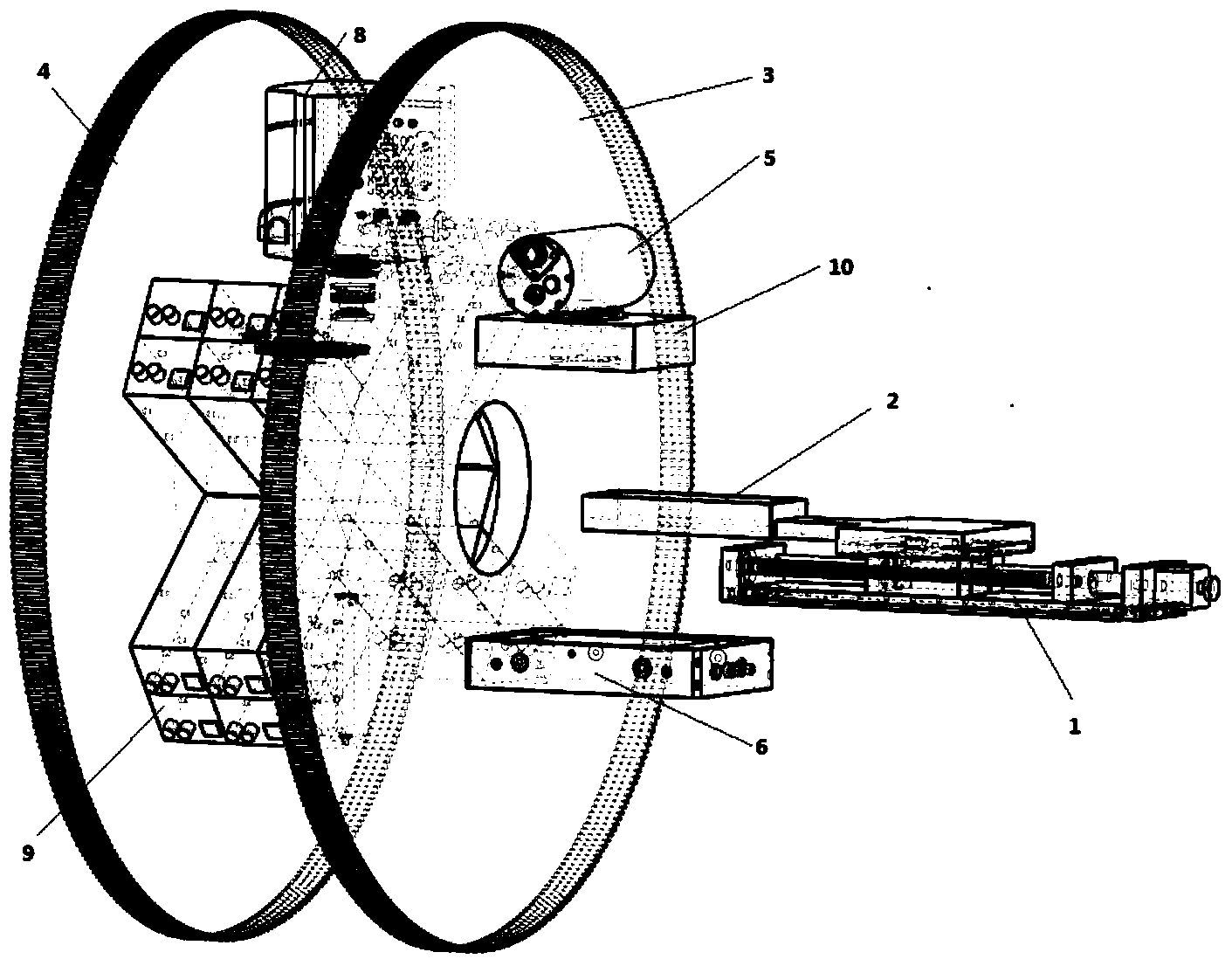
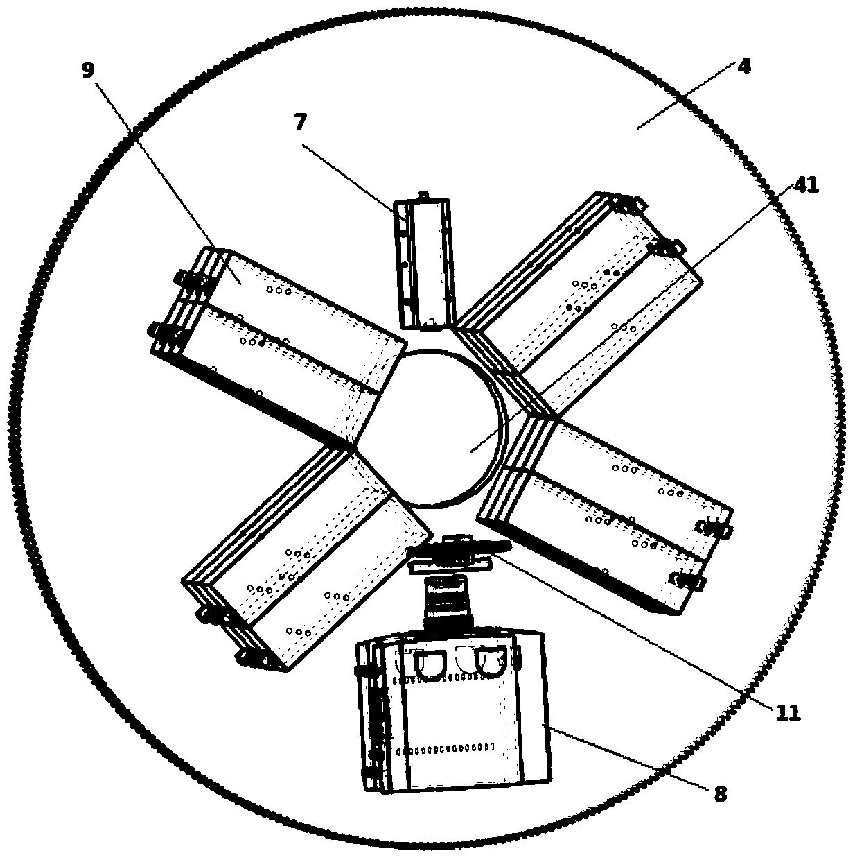
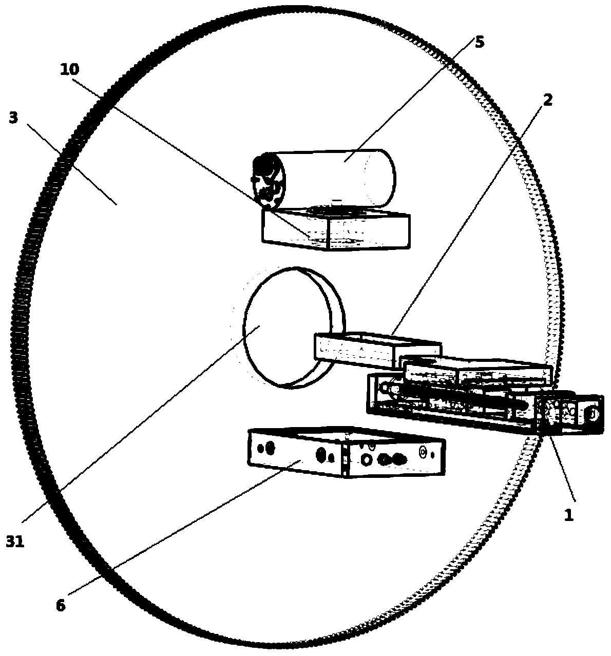
[0028] like Figures 1 to 4 As shown, the present invention is a CT / FT / PET three-modal synchronous imaging device, including small animals for observation, the device includes a translation platform 1 that can move back and forth horizontally, and a detection device connected to the translation platform 1 The bed 2, the CT frame 3 and the PET / FT frame 4, the CT frame 3 and the PET / FT frame 4 are provided with corresponding first imaging holes 31 and second imaging holes 41 to be The body axis of the small animal placed in the detection bed 2 is the central axis, and the centers of t...


PUM


Abstract

The invention discloses a CT/FT/PET three-mode synchronous imaging device which comprises a horizontally-moving table, a detection bed, a disc-shaped CT machine frame, a PET/FT machine frame, an X-ray tube, an X-ray detector, four PET detection heads, a laser, a CCD camera, an X-ray collimator, a filter wheel and a computer for constructing an image processing platform, wherein the disc-shaped CT machine frame and the PET/FT machine frame rotate with the axis of a detected small animal as the central axis, and each PET detection head is composed of six PET basic detection units. The CT/FT/PET three-mode synchronous imaging device integrates the advantages of the CT imaging mode, the FT imaging mode and the PET mode, more comprehensive and more accurate function and structure information can be provided for small animal carrier research, and the detection performance of early-stage tumors can be greatly improved.

Description

technical field [0001] The invention relates to the technical field of molecular imaging, in particular to a CT / FT / PET three-mode synchronous imaging device. Background technique [0002] Small animal in vivo research methods are widely used in various preclinical research fields such as tumor research and drug development, but small animal in vivo research must rely on imaging methods such as CT, PET, SPECT, and MRI. Single-modal imaging methods can only provide structural or functional images of small animals, which has great limitations. Therefore, the development of multi-modal imaging equipment has become a hot spot in the field of medical imaging equipment research. [0003] So far, several research teams and commercial institutions have developed a variety of multimodal systems for small animal research, such as Inveon PET / CT from Siemens Medical Devices, and GENISYS PET / CT commercialized by Sofie Biosciences developed by UCLA. In addition, there are dual-modal or tr...

Claims


Application Information

Patent Timeline
IPC IPC(8): A61B6/03A61B5/055
Inventor 田捷陈雪利刘红波朱守平曹旭陈多芳梁继民
Owner XIDIAN UNIV
Why Eureka
Try Eureka