Application of serum microRNA as early diagnostic marker for hepatocellular carcinoma metastasis

A technology of hepatocellular carcinoma, 1.microRNA-mir-107, applied in the fields of biotechnology and medicine, can solve problems such as large trauma, and achieve the effect of good sensitivity and specificity

Active Publication Date: 2014-07-23
青岛瑞思德医学检验实验室有限公司
View PDF11 Cites 4 Cited by
  • Summary
  • Abstract
  • Description
  • Claims
  • Application Information

AI Technical Summary

Problems solved by technology

(3) Non-invasiveness of detection
Although microRNA in tumor tissue can be used as a new marker for tumor diagnosi

Method used


Image

  • Application of serum microRNA as early diagnostic marker for hepatocellular carcinoma metastasis
  • Application of serum microRNA as early diagnostic marker for hepatocellular carcinoma metastasis
  • Application of serum microRNA as early diagnostic marker for hepatocellular carcinoma metastasis

Examples


Embodiment 1

[0047] Example 1 Serum Sample Collection

[0048] A total of 155 serum samples were collected, including 62 serum samples from patients with metastasis, 53 serum samples from patients without metastasis, and 40 healthy controls. Serum was collected from the Department of Infectious Diseases, Provincial Hospital of Shandong University. Serum was collected at the time of clinical diagnosis, from March 2012 to November 2013. Serum was collected in coagulation-promoting tubes, centrifuged at 2000-3000rpm for 18-20min, the supernatant was drawn into centrifuge tubes, and stored at -80°C. The clinical case characteristics of the patients are shown in Table 1.

[0049] Table 1 Clinical data of serum samples

[0050]

[0051]

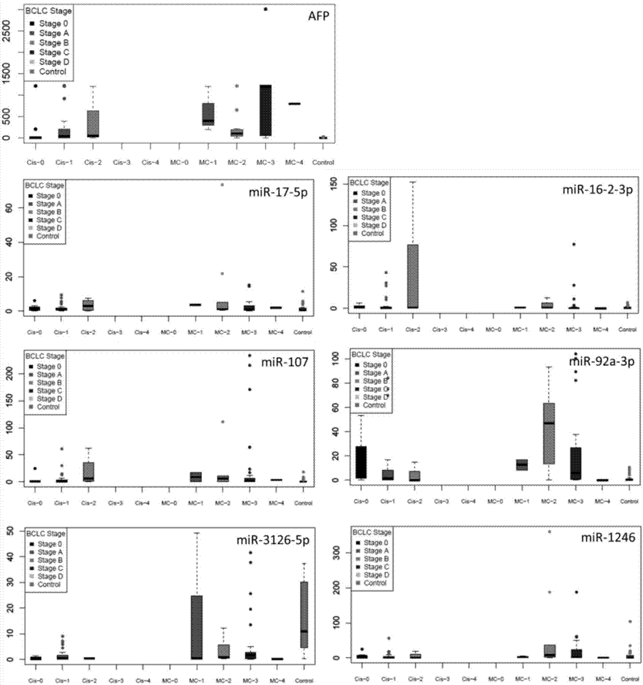
[0052] Tumor staging was according to the Barcelona classification system.

Embodiment 2

[0053] The detection of embodiment 2 microRNA chip

[0054] 1. Poly-HEMA plate preparation

[0055] Dissolve 2.4 g of Poly-HEMA powder in 20 mL of 95% ethanol, shake and mix at 65 °C for more than 8 h, prepare a Poly-HEMA stock solution with a concentration of 120 mg / mL, and store it in a sealed container at 4 °C. When preparing the plate, dilute it with 95% ethanol to form a working solution. Under sterile conditions, gently add the working solution to the bottom of the culture plate well along the well wall to cover the entire bottom of the well. Store at 4°C after ultraviolet irradiation overnight. Before use, it was irradiated under ultraviolet light for 0.5h and washed 3 times with sterile PBS.

[0056] 2. Preparation of liver cancer cell anoikis model

[0057] Liver cancer cell lines HepG2, BEL7402 and SMMC7721 were cultured in RPM11640 plus 10% fetal bovine serum at 37°C and 5% CO2 until logarithmic growth phase. Use 0.25% trypsin to digest the logarithmic phase adhe...

Embodiment 3

[0067] Example 3 Extraction of serum microRNA

[0068] (1) Take 400 μl of frozen patient serum, add 750 μl of lysate MRL, and pipette several times;

[0069] (2) Shake the homogenate sample vigorously, incubate at room temperature for 5 minutes, and carefully transfer the supernatant to a new RNasefree centrifuge tube.

[0070] (3) Add 200 μl chloroform for every 750 μl lysate, cover the sample tube tightly, shake vigorously for 15 seconds and incubate at room temperature for 3 minutes.

[0071] (4) Centrifuge at 12,000 rpm at 4°C for 10 minutes; the sample will be divided into 3 layers, remove the upper aqueous phase, and transfer to a new RNase freeEP tube.

[0072] (5) Add 0.6 times the volume of 70% ethanol, invert and mix well, and transfer the resulting solution and possible precipitates into the adsorption column RA.

[0073] (6) Centrifuge at 10000rpm for 45s, collect the filtrate, add 2 / 3 volume of absolute ethanol, mix by inversion, pour the mixture into adsorption...


PUM


Abstract

The invention discloses an application of miR-107 combined with miR-1246 as an early diagnostic marker for hepatocellular carcinoma metastasis. Researches find that miR-107 combined with miR-1246 for diagnosis of early liver cancer metastasis is superior to alpha fetal protein (AFP), and has good sensitivity and specificity, and the miR-107 and miR-1246 combined with AFP diagnosis is more excellent. As to metastasis diagnosis of low alpha fetal protein secretion liver cancer (AFP is smaller than 200), the miR-1246 is superior to the APF, the miR-107 combined with miR-1246 diagnosis is much superior to the APF, and the miR-107 and miR-1246 combined with AFP are more excellent. On the basis, a primer, a kit and a detection method for detecting the miR-107 and the miR-1246 are developed, and the serum microRNA has a good development prospect and clinical application value. A sequence of the primer is shown in SEQ ID NO.3 and 4, and the kit is formed by a specific amplification primer and a required reagent for PCR amplification.

Description

technical field [0001] The invention relates to the application of serum microRNA (miR-107 combined with miR-1246) as an early diagnosis marker for hepatocellular carcinoma metastasis, and belongs to the fields of biotechnology and medicine. Background technique [0002] Hepatocellular carcinoma (HCC) is the fifth most common malignant tumor of the digestive system in the world. It has the characteristics of high malignancy, high metastasis rate, high recurrence rate, and extremely poor prognosis. The morbidity and mortality are gradually increasing. , is the third leading cause of cancer-related deaths in the world. [0003] The recurrence rate of hepatocellular carcinoma is extremely high. According to domestic reports, the 5-year recurrence rate of patients with liver cancer after surgery is as high as 61.5%, and the 5-year survival rate is only 30% to 40%. The easy metastasis of liver cancer cells is an important factor causing the high recurrence rate after surgery, an...

Claims


Application Information

Patent Timeline
IPC IPC(8): G01N33/574
CPCG01N33/57438G01N33/57488G01N2800/50
Inventor 韩丽辉张颖李涛邱驭旻贾晓青赵伟怀婉婉郭蓬勃康佳姜艳艳
Owner 青岛瑞思德医学检验实验室有限公司
Why Eureka
Try Eureka