Monoclonal antibody against duck hepatitis A virus type A and application thereof

A duck hepatitis A virus, monoclonal antibody technology, applied in the direction of anti-virus immunoglobulin, biochemical equipment and methods, instruments, etc., to achieve the effect of good stability and strong specificity

Active Publication Date: 2014-09-10
SOUTHWEST UNIVERSITY FOR NATIONALITIES
View PDF2 Cites 6 Cited by
  • Summary
  • Abstract
  • Description
  • Claims
  • Application Information

AI Technical Summary

Problems solved by technology

However, there is currently no monoclonal antibody for genotype A DHAV detection and treatment

Method used


Image

  • Monoclonal antibody against duck hepatitis A virus type A and application thereof
  • Monoclonal antibody against duck hepatitis A virus type A and application thereof
  • Monoclonal antibody against duck hepatitis A virus type A and application thereof

Examples


Embodiment 1

[0023] In the process of peptide synthesis, a cysteine ​​residue is added to the C-terminal of the peptide, and the peptide fragment and KLH carrier protein are coupled through cysteine ​​with thermo's SMPH amphipathic peptide coupling reagent as an antigen.

[0024] Coupling process:

[0025] 1. Dissolve 20 mg of SMPH in 2 ml of DMF.

[0026] 2. Add 0.8 ml KLH to a 25 ml round bottom flask, add 1×PBS (pH 7.2) to make the final protein concentration 15 mg / ml.

[0027] 3. Slowly add the dissolved SMPH solution dropwise to the 120 mg KLH protein system, and stir at room temperature for 1 hour. 4. Dialyze with 1 L of 1×PBS (pH 7.4) solution at 4°C for 6 hours to remove free SMPH.

[0028] 5. Pour the dialyzed KLH protein into a 50 ml centrifuge tube, determine its volume through the scale of the centrifuge tube, calculate the concentration of the dialyzed protein according to the amount of KLH protein added before the reaction, and then add 2.5 mg KLH protein according to its c...

Embodiment 2

[0041] Method: The ascites titer of the monoclonal antibody obtained in step 1.4 was identified by indirect ELISA.

[0042] Results: The ascites titer of the monoclonal antibody of the present invention was 1:128 000.

[0043]2.2 Identification of monoclonal antibody activity

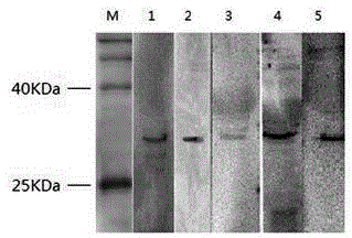
[0044] Methods: After SDS-PAGE electrophoresis, DHAV-A was semi-dryly transferred to PVDF membrane for Western blot analysis. The primary antibody was MAb diluted 1:200, and the secondary antibody was HRP-labeled goat anti-mouse IgG diluted 1:5000.

[0045] Results: The monoclonal antibody reacted specifically with DHAV-A virus, such as figure 1 , The hybridization results showed that the five monoclonal antibodies could hybridize with the virus and showed bands, indicating that the five monoclonal antibodies were active and could react with the antigen.

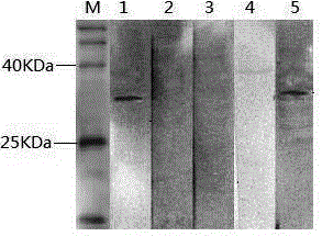
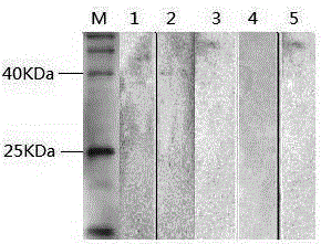
[0046] 2.3 Specific identification of monoclonal antibodies

[0047] Methods: DHAV-C, DPV, NDV and AIV were identified by SDS-PAGE electrophores...

Embodiment 3

[0054] DHAV-A clinical isolates were inoculated with 11-day-old duck embryos via the duck embryo allantoic cavity at a dose of 0.2 mL / embryo, discarded dead duck embryos within 24 hours, and collected dead and surviving duck embryo allantoic fluid 24-96 hours after inoculation , repeated freezing and thawing 3 times. Centrifuge at 12 000r / min at 4°C for 15 min, take the supernatant and mix it with an equal amount of chloroform; mix the above virus solution with an equal amount of chloroform, let stand at 4°C for 1 hour, centrifuge at 5,000r / min at 4°C for 30 min, and take the supernatant , repeat the above operation twice; take the supernatant and filter it with a 0.22 um filter and centrifuge at 40,000r / min in an ultracentrifuge for 2h, dissolve the precipitate with an appropriate amount of PBS buffer, and wash it through 20%, 30%, 40%, 50% A sucrose density gradient was centrifuged at 38,000r / min for 3h. Slowly absorb the suspension liquid at the obvious bright band in the ...


PUM


Abstract

The invention provides a monoclonal antibody against duck hepatitis A virus type A. According to the invention, mixed polypeptide is coupled with KLH carrier protein through cysteine residues so as to obtain an antigen which is used to immunize a mouse; spleen cells of the mouse are fused with SP2/0 bone marrow cells, and screening is carried out so as to obtain a hybridoma cell line 3L1-6 with a microbial accession number of CCTCC No: C2013134. The monoclonal antibody prepared from the hybridoma cell line has strong specificity and good stability and can be extensively used in preparation of detection reagents for antibodies against duck hepatitis A virus type A. An ELISA kit developed from the antibody has strong specificity and good stability and can be used for detection of antibodies against duck hepatitis A virus type A.

Description

technical field [0001] The invention relates to the field of molecular biology, in particular to a method for preparing a monoclonal antibody capable of treating duck hepatitis A virus and the use of preparing the antibody, and belongs to the field of prevention and control of gene A duck viral hepatitis. Background technique [0002] Duck viral hepatitis (DVH) is an acute and highly fatal infectious disease caused by duck hepatitis virus (DHV) that mainly harms ducklings under 3 weeks of age. DHV includes members of Picornaviridae and Astroviridae, and DHV of Picornaviridae is named as duck hepatitis A virus (DHAV). According to the difference in genotype structure, DHAV is divided into genotype A (DHAV-A), type B (DHAV-B) and type C (DHAV-C) three genotypes. Among them, the traditional type I DHV popular in my country is genotype A DHAV, and the new one in Taiwan is genotype B. In recent years, Korean-like DHV or genotype C has been widely distributed in my country. The c...

Claims


Application Information

Patent Timeline
Patent Type & Authority Applications(China)
IPC IPC(8): C07K16/10C12N5/20G01N33/577G01N33/569
Inventor 岳华汤承张焕容
Owner SOUTHWEST UNIVERSITY FOR NATIONALITIES
Why Eureka
Try Eureka