Three dimensional pixel super-resolution microscopic imaging method

A microscopic imaging and super-resolution technology, used in material excitation analysis, fluorescence/phosphorescence, etc., which can solve the problems of large field of view and high resolution that are difficult to be compatible.

Active Publication Date: 2014-10-22
HUAZHONG UNIV OF SCI & TECH
View PDF2 Cites 26 Cited by
  • Summary
  • Abstract
  • Description
  • Claims
  • Application Information

AI Technical Summary

Problems solved by technology

[0004] Aiming at the problem that the large field of view and high resolution are difficult to be compatible with the three-dimensional microscopic imaging technologies such as the above-mentioned inverted microscope, confocal fluorescence microscope, and sliced ​​light, the present invention provides a new three-dimensional pixel super-resolution microscopic imaging method

Method used


Image

  • Three dimensional pixel super-resolution microscopic imaging method
  • Three dimensional pixel super-resolution microscopic imaging method
  • Three dimensional pixel super-resolution microscopic imaging method

Examples


Embodiment Construction

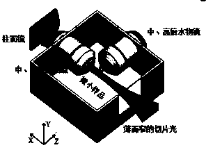
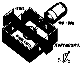
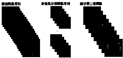
[0031] The three-dimensional structure of lung cells cultured in vitro is imaged by using the three-dimensional pixel super-resolution microscopic imaging method and slice light microscopic imaging technology of the present invention. Imaging settings were 4x acquisition objective + 10 µm thick illumination light sheet. According to this setting, in the obtained low-resolution three-dimensional image Bi, in the x-y direction, the size of a single pixel is the camera pixel size 6.45 μm / 4 = 1.61 μm, and in the z direction, the size of a single pixel is half the thickness of the light sheet, that is, 5 Micron. According to the Enquist sampling principle, the images correspond to raw lateral and axial resolutions of ~3.2 μm and ~10 μm, respectively. In the image acquisition, in order to achieve the super-resolution ratio of Ex=5, Ey=5, Ez=10, when generating 250 sets of low-resolution images, we selected an overscanning step size of 10000nm / 250=40nm. Next, the original image seq...


PUM


Abstract

A three dimensional pixel super-resolution microscopic imaging method is characterized by comprising the following steps: carrying out space vector scanning on a sample, wherein the scanning vector can form a non-right-angle spacial deflection angle with the plane horizontal direction X and vertical direction Y of an image collection device sensor or a Z axis direction of a microscope, so as to make that two neighbored image slices can be scanned by a step size of a sub pixel displacement along the X, Y, and Z directions; collecting the original three dimensional image sequence A by the image collecting device, cutting the original image sequence A into a plurality of groups of three dimensional image sequences Bi according to the lossless sampling principle, carrying out a super-resolution treatment on Bi to generate a three dimensional high resolution image E, and carrying out a deblurring treatment on E so as to obtain a clear high resolution image F. The spacial reflection angles formed by the scanning vector with the horizontal direction X, the vertical direction Y, and the Z axis of the microscope can be identical or different.

Description

technical field [0001] The invention relates to a microscopic imaging method, in particular to a three-dimensional pixel super-resolution microscopic imaging method. Background technique [0002] Fluorescence microscopy imaging, as a means of observing specific events in the microscopic world, has long been of great significance to modern life science research. Especially in developmental biology, regenerative medicine, tissue engineering and other fields that require the observation and analysis of multicellular samples, three-dimensional and all-round imaging is very important for analyzing the structure and biological characteristics of samples. However, neither the inverted microscope nor the confocal microscope can provide three-dimensional images with sufficient axial resolution under low magnification and large field of view. The newly developed slice light microscopic imaging system in recent years provides a new method of acquiring three-dimensional microscopic ima...

Claims


Application Information

Patent Timeline
Patent Type & Authority Applications(China)
IPC IPC(8): G01N21/64
Inventor 费鹏虞之龙董思炎关泽一严刚
Owner HUAZHONG UNIV OF SCI & TECH
Why Eureka
Try Eureka