Polypeptide-protein combined-type marker" detection kit relative to colorectal cancer

A technology of colorectal cancer and kits, applied in the field of biotechnology detection, to achieve good accuracy and sensitivity

Inactive Publication Date: 2014-11-19
BEIJING PROTEOME RES CENT +1
View PDF4 Cites 7 Cited by
  • Summary
  • Abstract
  • Description
  • Claims
  • Application Information

AI Technical Summary

Problems solved by technology

[0006] However, there is still little research on the new "polypeptide-protein combined marker" at present. Th

Method used


Image

  • Polypeptide-protein combined-type marker" detection kit relative to colorectal cancer
  • Polypeptide-protein combined-type marker" detection kit relative to colorectal cancer
  • Polypeptide-protein combined-type marker" detection kit relative to colorectal cancer

Examples


Embodiment 1

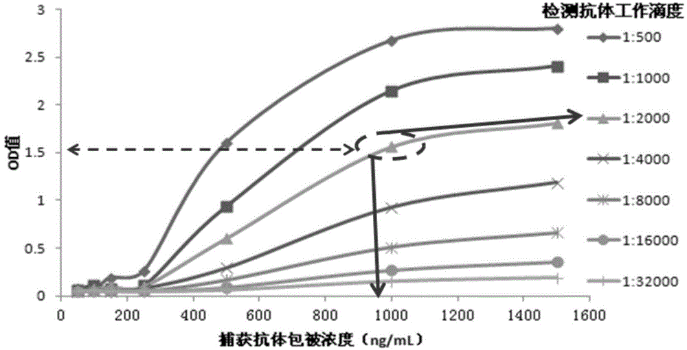
[0071] Example 1: Optimization of Detection Conditions for Capture Antibodies and Detection Antibodies

[0072] 1) Dilute the capture antibody with coating buffer to 50ng / mL, 100ng / mL, 250ng / mL, 500ng / mL, 1000ng / mL and 2000ng / mL, respectively, according to 100μL / well, and place overnight at 4°C.

[0073] 2) Take the ELISA plate coated with the capture antibody in the kit, add 100 ng / mL HKa standard to each well, 100 μL per well. Incubate at 37°C for 1.5 hours;

[0074] 3) Discard the solution, wash each well 5 times with 300 μL PBST washing buffer, and pat dry;

[0075] 4) Dilute the HRP-labeled detection antibody with PBST at 1:500, 1:1000, 1:2000, 1:4000, 1:8000, 1:16000, 1:32000, add 100 μL to each well, and stand at 37°C Incubate for 40 minutes;

[0076] 5) Discard the solution, wash each well with 300 μL PBST washing buffer 5 times, and pat dry;

[0077] 6) Add 100 μL of TMB substrate chromogenic solution interacting with HRP to each well, and develop color at 37°C in...

Embodiment 2

[0080] Embodiment 2: the preparation of kit

[0081] 1. Preparation of reagents

[0082] Carbonate coating buffer: pH9.6, 0.1mol / L, weighed Na 2 CO 3 1.59g, NaHCO 3 Add 2.93g of deionized water to 1000ml.

[0083] PBST washing buffer: pH value is 7.4, including 80mmol / L Na 2 HPO 4 , KH of 20mmol / L 2 PO 4 , KCl of 100mmol / L, NaCl of 1400mmol / L, Tween-20 whose volume fraction is 0.5%.

[0084] TMB Chromogenic Solution: Substrate Chromogenic Solution A: Sodium Acetate 13.6g, Citric Acid 1.6g, 30% Hydrogen Peroxide 0.3ml, Distilled Water to 500ml; Substrate Chromogenic Solution B: Disodium EDTA 0.2g, Citric acid 0.95g, glycerin 50ml, 0.15g TMB was dissolved in 3ml DMSO, and distilled water was added to 500mL. Take substrate color development solution A and mix equal volumes of substrate color development solution B to obtain TMB color development solution.

[0085] Sulfuric acid stop solution: prepare a 2M sulfuric acid aqueous solution.

[0086] 2. Coating of capture...

Embodiment 3

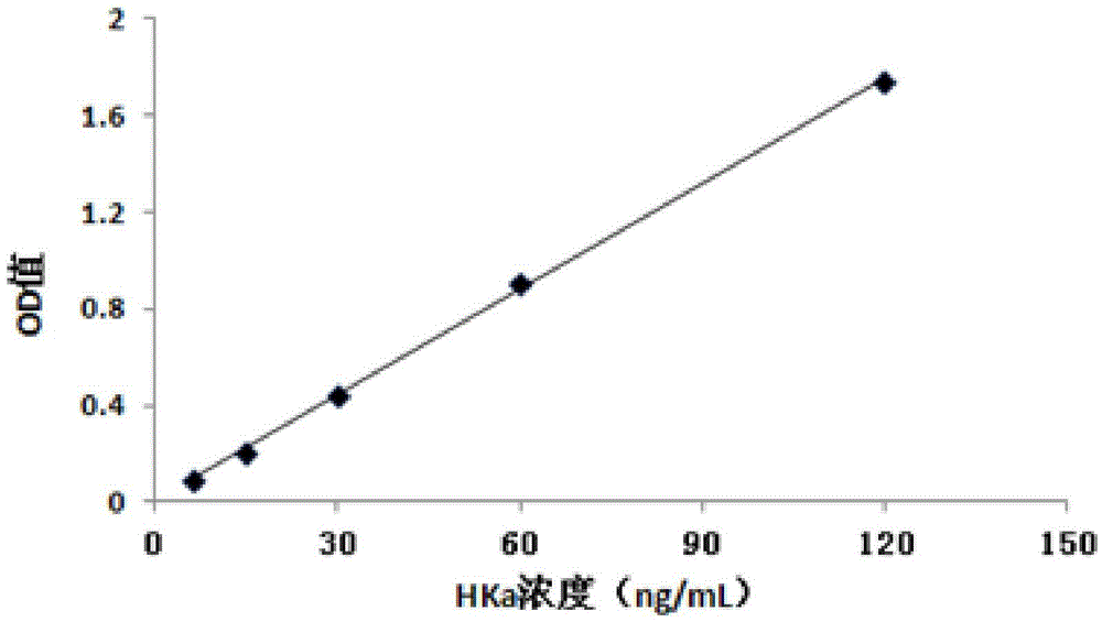
[0092] Example 3: Identification of the minimum detection limit and detection range of the kit

[0093] Get the kit that embodiment 2 makes, prepare the HKa standard solution of gradient concentration as the sample to be tested, the concentration of HKa standard is: 0ng / mL, 30ng / mL, 60ng / mL, 90ng / mL, 120ng / mL, 150ng / mL.

[0094] Take the ELISA plate coated with the capture antibody in the kit, add different concentrations of HKa standard solution, 100 μL per well, and repeat 3 times for each concentration. Incubate at 37°C for 1.5 hours; discard the solution, wash each well 5 times with 300 μL PBST buffer in the kit, and pat dry;

[0095] Add 100 μL of HRP-labeled detection antibody to each well, and incubate at 37°C for 40 minutes; discard the solution, wash each well 5 times with 300 μL of PBST buffer in the kit, and pat dry;

[0096] Add 100 μL of TMB chromogenic solution to each well, and develop color at 37°C in the dark for 15 minutes; add 50 μL of 2mol / L sulfuric acid...


PUM


Abstract

The invention relates to the technical field of biologic and especially relates to a "polypeptide-protein combined-type marker" detection kit relative to colorectal cancer. The kit contains a capture antibody and a detection antibody, wherein the capture antibody is specifically combined to protein HKa and the detection antibody is specifically combined to polypeptide HKP15 or polypeptide HKP09. To a HKP09/HKP15-HKa combined-type marker, the kit has a linear detection range being 6-120 ng/ml and a minimum detection limit being 1.7 ng/ml. To distinguish a colorectal cancer sample from a normal human sample, the kit is 93.7% in sensitivity and is 92.7% in specificity. The novel combined marker detected in the kit provided in the invention is closely relative to the colorectal cancer. In addition, a detection technology is very high in accuracy and sensitivity, is simple and quick in operation, is beneficial to high-flux clinical detection and can reduce a detection cost.

Description

technical field [0001] The invention relates to the field of biotechnology detection, in particular to a colorectal cancer-related "polypeptide-protein combined marker" detection kit. Background technique [0002] Colorectal cancer (Colorectal Carcinoma, referred to as CRC), including colon cancer and rectal cancer, is a common malignant tumor of the digestive tract and has a high incidence in economically developed countries such as North America, Western Europe, Australia, New Zealand and other countries. In countries and regions with rapid economic growth, the incidence rate is also rising rapidly. Therefore, the early diagnosis of colorectal cancer and the discovery of related diagnostic markers are urgent problems to be solved. [0003] Common screening methods for colorectal cancer include fecal occult blood test, sigmoidoscopy, total colonoscopy, and barium enema. At present, the main diagnostic methods are colonoscopy, CT simulation scanning, and pathological slide...

Claims


Application Information

Patent Timeline
IPC IPC(8): G01N33/68
CPCG01N33/57419
Inventor 魏开华侯利平孙云波宋纯艳杨保安周婷婷甄蓓刘曙晨郑俊杰
Owner BEIJING PROTEOME RES CENT
Why Eureka
Try Eureka