
Hemostatic Vacuum Device and Hemostatic Vacuum Scalpel

A vacuum device and scalpel technology, applied in the field of medical devices, can solve the problems of loose scabs, human body irritation, secondary bleeding, etc., and achieve the effects of reducing the risk of secondary bleeding, convenient operation and use, and stable coagulation.

Inactive Publication Date: 2016-10-19
3M INNOVATIVE PROPERTIES CO
View PDF4 Cites 0 Cited by
  • Summary
  • Abstract
  • Description
  • Claims
  • Application Information

AI Technical Summary

Problems solved by technology

Most of the existing hemostatic agents come from biological sources, such as collagen (gelatin or collagen), cellulose, chitosan, starch, fibrin, etc., which may have the potential risk of causing irritation, allergy and inflammation in the human body
Suture blood vessel hemostasis is usually used to deal with relatively large blood vessels, which is difficult to apply to capillary hemostasis
Surgical electric knife hemostasis is usually used to sinter the tissue at the bleeding site, but the sintered tissue may have potential risks of infirm scabbing and unstable coagulation, which may cause secondary bleeding after surgery

Method used


Image

  • Hemostatic Vacuum Device and Hemostatic Vacuum Scalpel
  • Hemostatic Vacuum Device and Hemostatic Vacuum Scalpel
  • Hemostatic Vacuum Device and Hemostatic Vacuum Scalpel

Examples


Embodiment 1

[0068] In this test, the rabbit extracorporeal thromboelastography test is mainly carried out using a thromboelastometer and rabbit whole blood. The thromboelastometer can be a TEG5000 thromboelastometer produced by American Blood Technology Corporation.

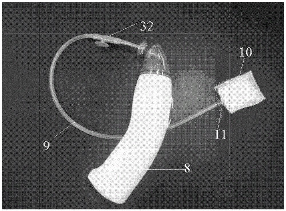
[0069] In this experiment, the hemostatic vacuum device provided by the present invention includes a vacuum generating device, a connecting device and a semi-permeable membrane. The vacuum generating device is a portable sputum suction device (model 7E-A / B) produced by Jiangsu Yuyue Medical Equipment Co., Ltd., and the vacuum generating device can provide a negative pressure with a vacuum degree of 0.01Mpa. The connecting device is a suction tube of the portable sputum suction device, and the semi-permeable membrane covers the tip of the suction tube. The semi-permeable membrane may be a semi-permeable membrane produced by Sefar Company, the material of the semi-permeable membrane is nylon, and the pore size is 1 μm.

[0070] Th...

Embodiment 2

[0085] Place a rabbit weighing about 2kg on its back on the operating table and separate the two hind legs; cut off the rabbit’s skin and cut the muscle to expose and isolate the femoral artery; use surgical scissors to cut the femoral artery to produce bleeding (The incision wound is about one-third the thickness of the femoral artery). The semipermeable tip of the hemostatic vacuum scalpel provided by the present invention is placed on the wound site, and a force of 50 g (about 0.49 N) is applied. The semi-permeable tip is made of a semi-permeable membrane, the semi-permeable membrane is a semi-permeable membrane made of nylon purchased from Sefar Company, the pores of the semi-permeable membrane are arranged regularly, and the semi-permeable membrane The diameter of the hole is 1 μm. The hemostatic vacuum scalpel includes a suction tube, one end of the suction tube is connected to the vacuum generating device, the other end of the suction tube is connected to the semipermea...

Embodiment 3

[0090] The rabbit femoral artery incision test was performed in the same way as in Example 2, except that the diameter of the semipermeable membrane hole was 5 μm.

[0091] List the test results in Table 2 and Figure 7 , Each test result is the average of the hemostasis time of six tests.


PUM


Abstract

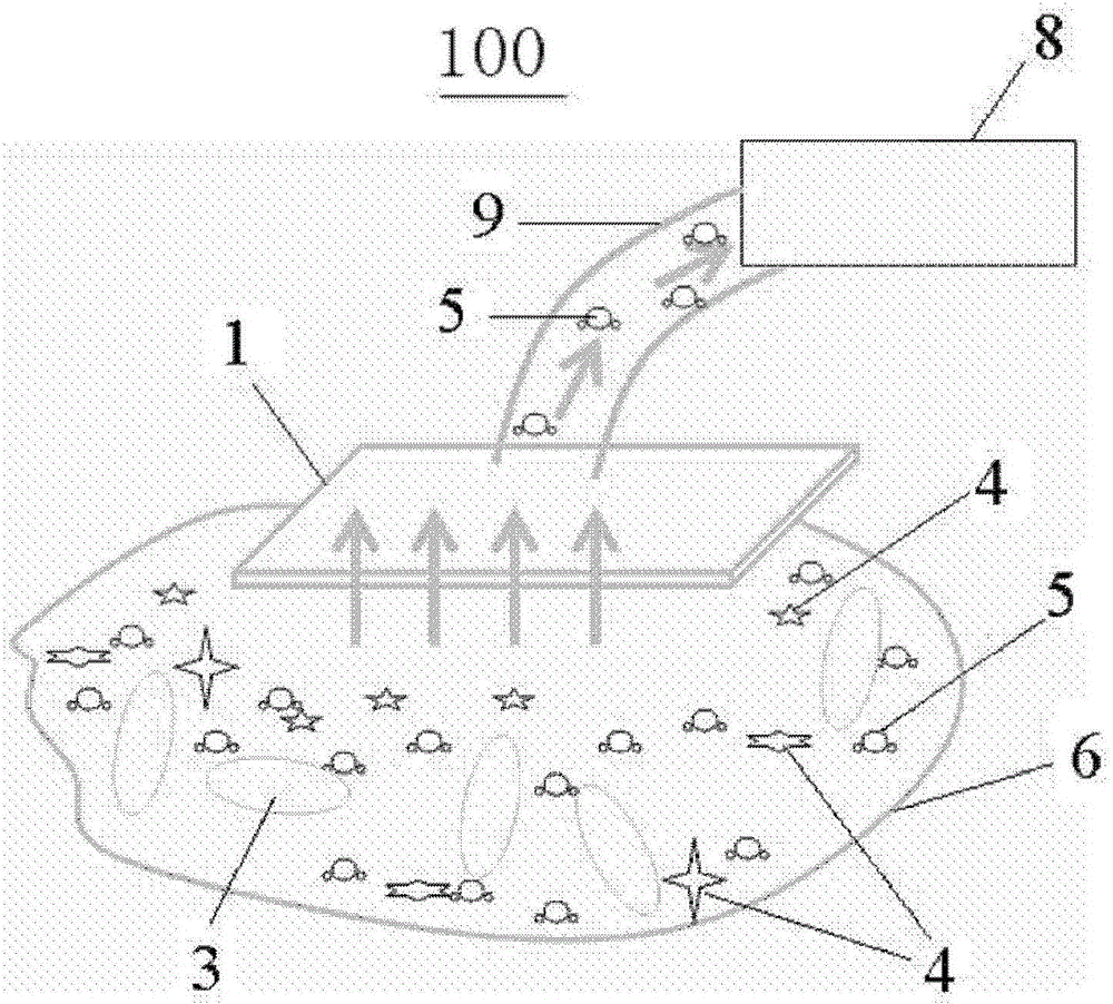
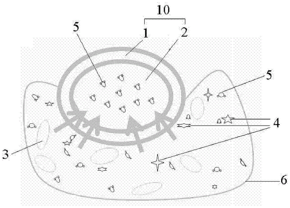
The invention provides a hemostasis vacuum device and a hemostasis vacuum scalpel. The hemostasis vacuum device comprises a vacuum generating device, a connecting device with one end connected to the vacuum generating device, and a semi-permeable membrane which is provided with holes for blocking blood platelets and / or blood cells and allowing water molecules to pass through. The other end of the connecting device is connected to the semi-permeable membrane. Thus, the hemostasis vacuum device can absorb moisture in blood at a bleeding part through the holes of the semi-permeable membrane so that the effect of accelerating the hemostasis can be achieved.

Description

Technical field [0001] The present invention relates to the field of medical equipment, in particular to a hemostatic vacuum device and a hemostatic vacuum scalpel used to stop bleeding during surgery. Background technique [0002] Usually, hemostasis is the first step in wound treatment. Especially in surgery, hemostasis is a particularly important step. [0003] At present, there are mainly the following methods of hemostasis in surgery: hemostasis with hemostatic agents, hemostasis with suture of blood vessels and hemostasis with electrosurgical knife. Most of the existing hemostatic agents come from biological sources, such as collagen (gelatin or collagen), cellulose, chitosan, starch, fibrin, etc. They may have potential risks of causing human irritation, allergies and inflammation. Suture blood vessel hemostasis is usually used to treat relatively large blood vessels, and it is difficult to apply capillary hemostasis. The electrosurgical method of hemostasis is usually us...

Claims


Application Information

Patent Timeline
Patent Type & Authority Patents(China)
IPC IPC(8): A61B17/12A61M1/00
CPCA61B17/12A61B2017/12004A61M1/0023
Inventor 刘恺鹏龚祖光范婷刘军钪刘杰白峰吴平凡
Owner 3M INNOVATIVE PROPERTIES CO
Learn More