Test bar and method for quantitatively detecting micromolecular compound in sample
A technology for small molecule compounds and test strips, applied in the field of detection, can solve the problems affecting the accuracy of test results and the high background of detection
- Summary
- Abstract
- Description
- Claims
- Application Information
AI Technical Summary
Problems solved by technology
Method used
Image

Examples
Embodiment 1
[0090] Preparation of fluorescent latex-labeled ketamine antibody (antibody complex)
[0091] Take 100 μl of 10% europium latex microspheres with activated carboxyl groups on the surface (200 nm, purchased from Thermo Fisher, USA), add 900 μl of MES (0.05M, pH6.1) to wash, centrifuge at 13000 rpm for 15 minutes, and discard the supernatant. Add 1ml marker solution (0.05M MES pH6.1, 2.0mg / ml N-hydroxysuccinimide NHS, and 0.5mg / ml EDC.HCl (1-ethyl-3-(3-dimethylaminopropyl )-carbodiimide EDC)) resuspended, sonicated, and reacted at room temperature for 1 hour. The activated latex was centrifuged at 13,000 rpm for 15 minutes, and the supernatant was discarded. Resuspend with 1ml 0.05M MES buffer pH6.1. After sonication, 80 μg / ml of ketamine antibody was added, mixed evenly, and reacted at room temperature for 2 hours. The labeled latex was centrifuged at 13,000 rpm for 15 minutes, and the supernatant was discarded. Add 1ml of 0.05M MES buffer pH6.14%BSA to block, sonicate, and...
Embodiment 2
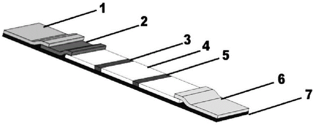
[0093] Preparation of immunochromatographic detection reagent plate
[0094] (1) Preparation of binding pad (marker pad)
[0095] The antibody complex and mouse IgG prepared in Example 1 were mixed with 10 mM phosphate buffer solution at pH 7.4 containing 0.5% surfactant Tetronic-1037 (purchased from BASF) to prepare a solution with a concentration of 0.5 mg / ml. Coat evenly on glass cellulose paper or polyester film, the coating amount is 50μl / cm 2 , vacuum dried. A conjugated pad is prepared.
[0096] (2) Detection area and quality control area
[0097] Detection area (also known as detection line): Dissolve ketamine bovine serum albumin (BSA) conjugate in 0.01M pH7.3 phosphate buffer containing 10% sucrose, spray on the nitrocellulose reaction membrane, Coating amount is 10μl / cm 2 ; Then dry at 15-35°C for 20 hours to make a detection area;
[0098] Quality control area (also called control line or control area): Dissolve goat anti-mouse IgG in 0.01M pH7.3 phosphate bu...
Embodiment 3
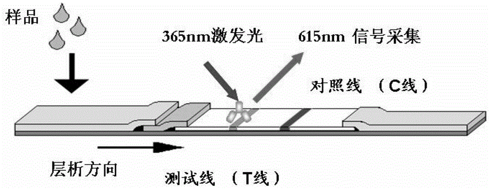
[0111] detection
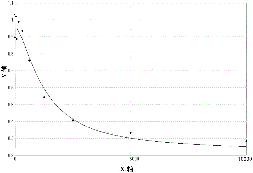
[0112] (1) Draw a standard curve
[0113] Prepare 1.0ml of ketamine solution with a concentration of 1000ng / ml in PBS, take 500μl and dilute with PBS to obtain different concentrations of ketamine solutions, as follows: 10000ng / ml, 5000ng / ml, 2500ng / ml, 1250ng / ml, 625ng / ml , 312.5ng / ml, 156ng / ml, 78ng / ml, 39ng / ml, 0ng / ml. PBS was used as blank control. Take 200 μl of sample and add it directly to the sample pad, react at room temperature for 5 minutes, then insert the test strip into the detection device for detection, obtain the fluorescence values of the T line in the detection area and the C line in the quality control area, and calculate the T / C ratio. The results are as follows Table 1 shows.
[0114] Table 1 Fluorescence detection data
[0115] Ketamine concentration (ng / ml)
T-line fluorescence value
C-line fluorescence value
T / C ratio
10000
5017
17724
0.283063
5000
5914
17730
0.333559
...
PUM
| Property | Measurement | Unit |
|---|---|---|
| molecular weight | aaaaa | aaaaa |
| emission peak | aaaaa | aaaaa |
| Sensitivity | aaaaa | aaaaa |
Abstract
Description
Claims
Application Information