Tumor specific target and application thereof in preparing preparation for cellular immunotherapy
A tumor-specific, cellular immune technology, applied in animal cells, anti-tumor drugs, vertebrate cells, etc., can solve problems such as staying
- Summary
- Abstract
- Description
- Claims
- Application Information
AI Technical Summary
Problems solved by technology
Method used
Image
Examples
Embodiment 1
[0018] Example 1: In vitro amplification and activation method for tumor-specific dendritic cell (DC) targets
[0019] 1. Specimen collection:
[0020] The blood picture of melanoma patients is required to be within the normal range (WBC: 4-10×10 9 , LYM%: 20%-40%), the fluctuation does not exceed 5%, the circulation volume of peripheral blood by machine sampling is 1000-4000ml, the number of mononuclear cells: more than 1×10 9 . Blood drawing method 50-100ml, the number of mononuclear cells: more than 1×10 6 .
[0021] The preferred anticoagulant is heparin, followed by sodium citrate, and EDTA is prohibited.
[0022] The collected specimens should not be stored for more than 6 hours, and cell preparation should be performed as soon as possible. If the specimen is stored for more than 2 hours before preparation, the specimen should be stored at 4°C.
[0023] 2. Preparation before operation
[0024] Wipe the collection bag containing the suspension of PBMC peripheral bl...
Embodiment 2
[0055] Example 2: Test method for detection of cell positive rate by target monoclonal antibody
[0056] Positive detection steps of indirect immunofluorescence on the surface of peripheral blood DC cells in patients with colon cancer
[0057] 1. Take the peripheral blood DC cells (1×10 7 / ml) 200 μl, divided into two tubes, one tube for each experimental control (100 μl / tube).
[0058] 2. Centrifuge and wash once with PBA,
[0059] 3. Add 20ul of the target monoclonal antibody diluted with PBA to the experimental tube, add 20ul of PBA to the control tube, gently pipette and mix, and incubate at 4°C or on ice for 1.5-2h. Centrifuge and discard the supernatant.
[0060] 4. Centrifuge and wash once with 1ml of PBA to remove excess unbound specific antibody.
[0061] 5. 20ul of fluorescein-labeled secondary antibody appropriately diluted in PBA. Mix by pipetting and incubate at 4°C for 30 min, protected from light. (PBA: PBS plus 1-2% bovine serum albumin, plus 0,1% sodium ...
Embodiment 3
[0066] Embodiment 3: the test method of DC cell marker antibody detection cell positive rate
[0067] Positive detection steps of indirect immunofluorescence on the surface of peripheral blood DC cells in patients with melanoma
[0068] 1. Take the peripheral blood DC cells (1×10 7 / ml) 200 μl, divided into two tubes, one tube for each experimental control (100 μl / tube).
[0069] 2. Centrifuge and wash once with PBA;
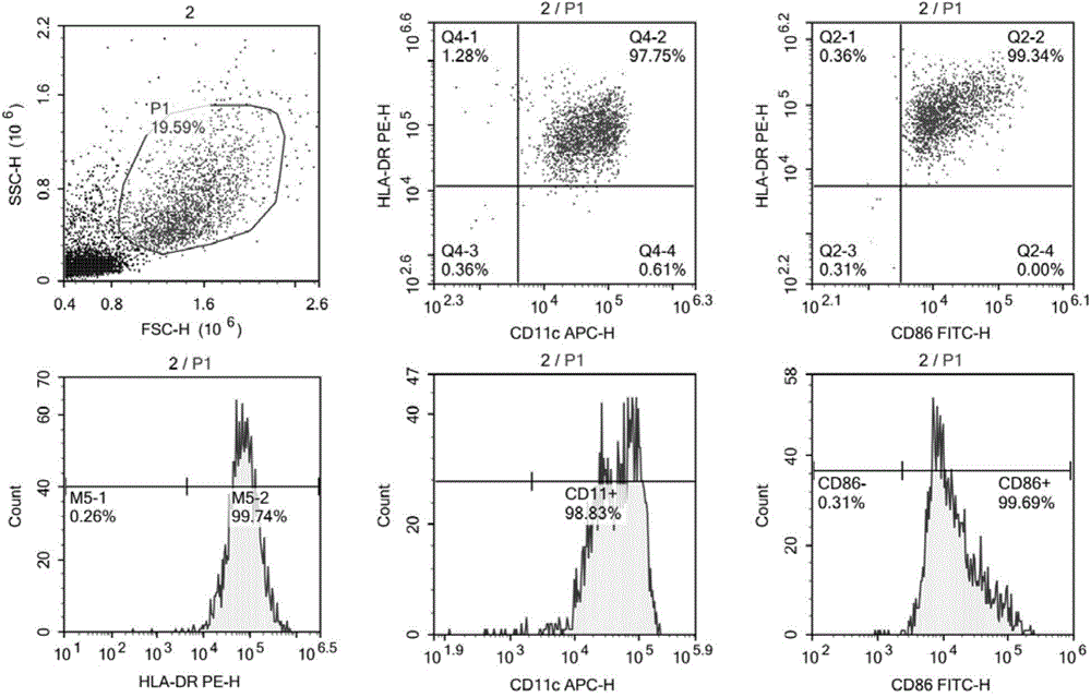
[0070] 3. Add 20 μl each of FITC-CD86, PE-HLA-DR, and APC-CD11c to the experimental tube, and add 20 μl each of FITC-mouse IgG1, PE-mouse IgG1, and APC-mouse IgG1 to the control tube.
[0071] 4. Gently blow and beat to mix well. After each tube is fully mixed, incubate in the dark for 30 minutes.
[0072] 5. After washing with PBS, test on the machine.
[0073] 6. See the test results image 3 ,analyse as below:
[0074] Experimental tube:
[0075] HLA-DR + Cells: 99.74%
[0076] CD11c + Cells: 98.83%
[0077] CD86 + Cells: 99.69%
[0078] HLA-DR ...
PUM
 Abstract
Description
Claims
Application Information
 - R&D
- Intellectual Property
- Life Sciences
- Materials
- Tech Scout
- Unparalleled Data Quality
- Higher Quality Content
- 60% Fewer Hallucinations
Browse by: Latest US Patents, China's latest patents, Technical Efficacy Thesaurus, Application Domain, Technology Topic, Popular Technical Reports.
© 2025 PatSnap. All rights reserved.Legal|Privacy policy|Modern Slavery Act Transparency Statement|Sitemap|About US| Contact US: help@patsnap.com


