Rationality analyzing method for blood vessel operation mode

An analytical method, a technique for vascular surgery, applied in the fields of surgery and computer crossover, physiology, and pathophysiology, which can solve the problems of high cost, interference, and lack of information about blood flow

Inactive Publication Date: 2015-06-24
THE FIRST AFFILIATED HOSPITAL OF SUN YAT SEN UNIV
View PDF0 Cites 9 Cited by
  • Summary
  • Abstract
  • Description
  • Claims
  • Application Information

AI Technical Summary

Problems solved by technology

In addition, this method needs to be completed under the guidance of X-ray fluoroscopy, and both the patient and the doctor will receive radiation; this technique is difficult to operate, and complications such as thrombosis and puncture of the artery may occur
[0006] Second, postoperative non-invasive detection
However, this method is highly subjective, related to the technique of the detector, and is also interfered by many factors.
[0008] 2. CT angiography: Its advantage is that it can provide rich anatomical information, but its disadvantage is that it can only provide information on anatomical structures and lack information on blood flow
However, the currently applied technology cannot quantify the information on the interaction between blood and blood vessel walls such as pressure indicators, and its high price also limits its clinical application to a certain extent.
[0010] To sum up, the current commonly used methods can only analyze the situation after vascular anastomosis, and can only analyze the simple flow rate and flow rate of blood flow.

Method used


Image

  • Rationality analyzing method for blood vessel operation mode
  • Rationality analyzing method for blood vessel operation mode
  • Rationality analyzing method for blood vessel operation mode

Examples


Embodiment 1

[0059] Embodiment 1 establishes the original 3D model

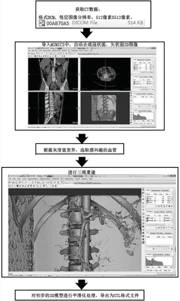
[0060] The enhanced CT images collected by the medical imaging work platform were exported to DCM format, with a layer thickness of 1.250 mm, a total of 1200 layers, an image resolution of 512 pixels by 512 pixels, and a pixel size of 0.797 mm. The picture is imported into MIMICS, and the software automatically calculates and generates a two-dimensional map (coronal plane, sagittal plane) composite map of the arterial system. According to the gray value difference between the blood vessel and the surrounding tissue, set the appropriate threshold, then limit the region of interest (ie artery), remove the non-arterial selection area, calculate the preliminary 3D model, use the smoothing function, and finally export it as STL format file.

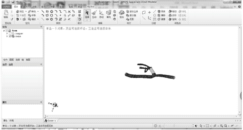
[0061] Import the STL format file into Ansys-SCDM, cut out the required arterial segment by establishing a plane, delete unnecessary structures, fill in the concave and convex parts of t...

Embodiment 2

[0062] Embodiment 2 rebuilds the original 3D model

[0063] Import the SCDOC format file in Example 1, cut and separate the artery to be reconstructed from the original artery, construct the vessel segment after the blood vessel is placed in the stent, obtain the reconstructed 3D model, and save it as a SCDOC format file. The reason for the reconstruction of the artery is to simulate different surgical methods.

Embodiment 3

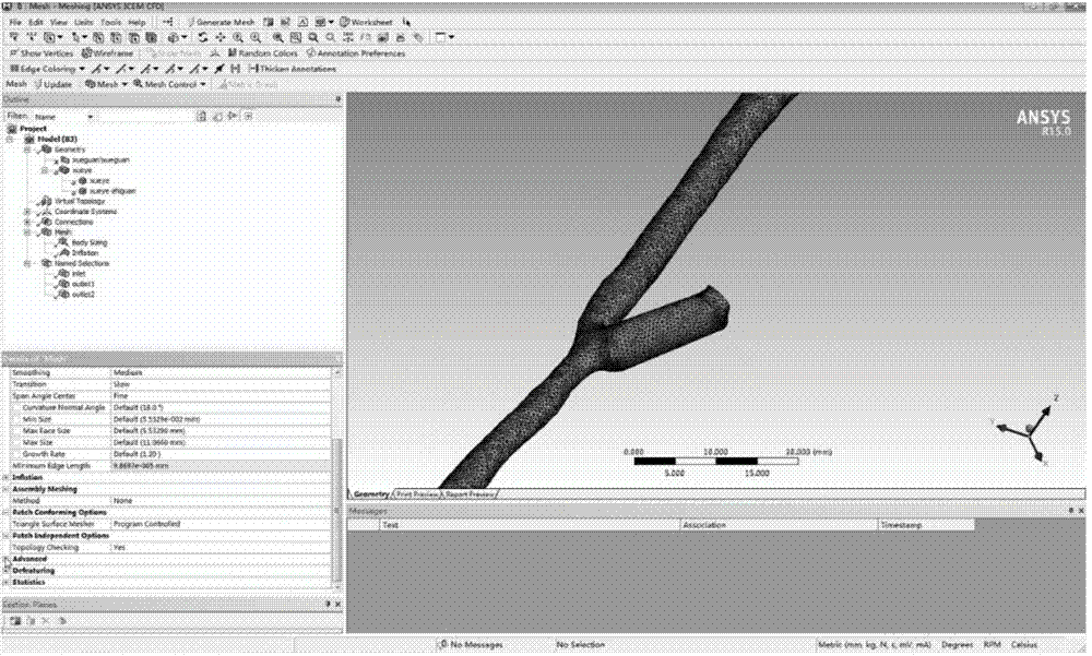
[0064] Embodiment 3 Gridded 3D model and parameter setting before simulation

[0065] Import the model into the mesh software for meshing. Adjust the size to 0.5mm; encrypt the grid near the blood vessel wall, and set it to 5 layers through inflation; check the grid quality, the skewness is 0.95, and the grid is acceptable. Then, set initial conditions such as material properties (blood density 1060kg / m 3 , the blood viscosity is realized by writing UDF using the Carreau model, and the inlet velocity equation is:

[0066] v n ( · , t ) = v ‾ ( t ) × n + 2 n ( 1 - ( r R ...


PUM


Abstract

The invention discloses a rationality analyzing method for a blood vessel operation mode. The analyzing method comprises the following steps that 1, artery CT image data are obtained to set up an artery 3D model; 2, rebuilding is carried out on the basis of the 3D model; 3, MESH software is used for meshing 3D models obtained in the step 1 and the step 2 to obtain a mesh 3D model capable of being used for calculation; 4, FLUENT software is used for simulating and calculating blood flow data of the artery to obtain parameters of WSS, OSI and blood vessel wall deformation. The method is safe, noninvasive and easy to operate, the intuitive quantized reference is provided for selecting vascular anastomosis modes, and the effective model for scientific research and clinic service is set up.

Description

technical field [0001] The invention relates to the interdisciplinary fields of physiology, pathophysiology, surgery and computer, and belongs to the analysis of surgical methods for vascular diseases, and specifically analyzes the rationality of the vascular surgical methods by measuring the parameters of blood's action on the vessel wall. Background technique [0002] In the current surgical operation, there is still a lack of a standard for the selection of surgical reconstruction methods for vascular diseases and surgical practice. However, the commonly used detection means can only perform simple flow velocity and flow detection after the operation. They mainly include invasive detection (such as digital subtraction angiography) and noninvasive detection (color Doppler ultrasound, CT angiography, magnetic resonance angiography). [0003] 1. About postoperative invasive testing [0004] 1. Digital subtraction angiography: This method is considered to be the "gold stand...

Claims


Application Information

Patent Timeline
Patent Type & Authority Applications(China)
IPC IPC(8): A61B19/00G06F17/50
CPCG06F30/20
Inventor 费继光曹海明王洪阳李军傅茜熊韫袆王长希
Owner THE FIRST AFFILIATED HOSPITAL OF SUN YAT SEN UNIV
Why Eureka
Try Eureka