Left ventricle volume reduction device
A technology of left ventricle and main body, which is applied in the field of left ventricle volume reduction device, can solve the problems that the position cannot reach the optimal position, the doctor’s operation requirements are relatively high, and the success rate of surgery is low, so as to reduce the risk of surgery and complications, and reduce the risk of surgery. Risk, effect of simplified operation
- Summary
- Abstract
- Description
- Claims
- Application Information
AI Technical Summary
Problems solved by technology
Method used
Image
Examples
Embodiment 1
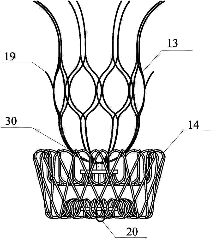
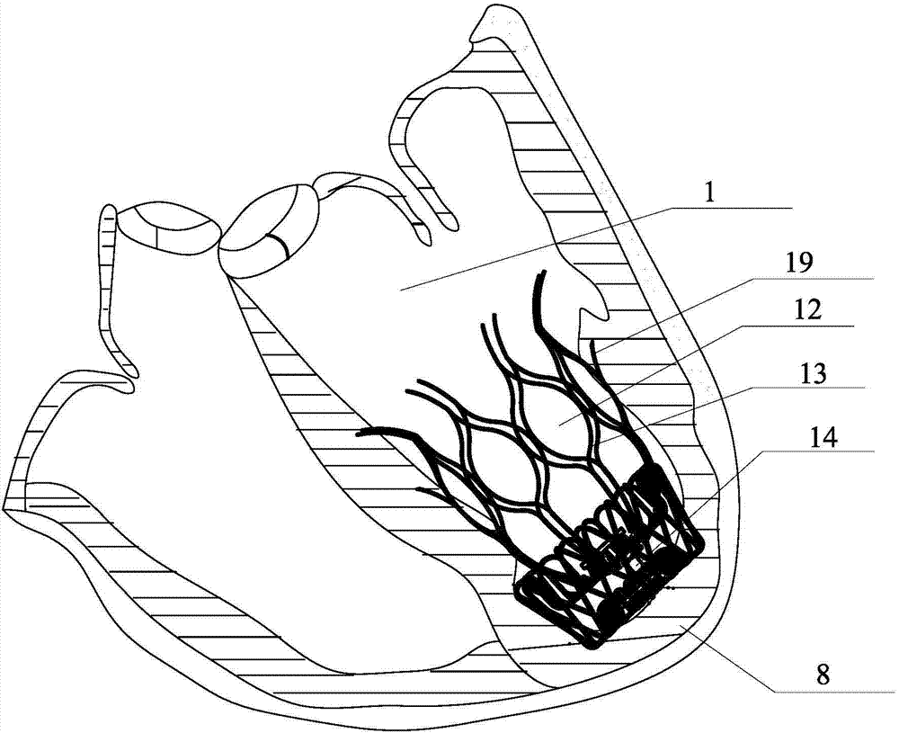
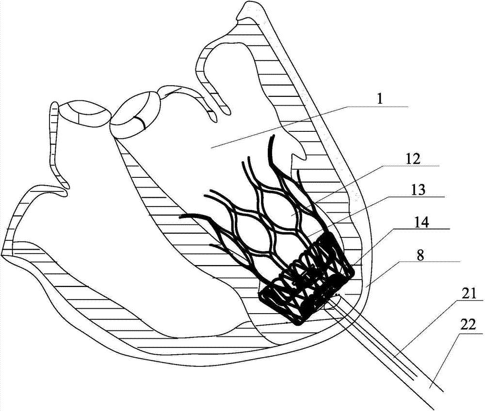
[0039] Such as figure 1 , 2 As shown, the left ventricular volume reduction device of the present invention adopts a two-stage design, which are the main body 13 and the base 14 respectively. The main body 13 is integrally formed by laser cutting of a nickel-titanium alloy tube, and the nickel-titanium alloy tube used has anti-magnetic properties and does not affect nuclear magnetic resonance imaging. Its cage-like structure has excellent mechanical stability, is not easy to deform, break and fall apart, and maintains its structural integrity. Such as figure 2 As shown, the left ventricular volume reduction device involved in the present invention can be placed in the left ventricle 1 through the aorta or through the apex, the base 14 is located at the position of the left apex 8, and the main body 13 is attached to the outer wall of the left ventricle 1 and the interventricular septum 3 Placed above the base 14, isolate the inactivated myocardium below the left ventricle ...
Embodiment 2
[0042] Such as Figure 5 , 6 , 7a, 7b, and 7c, this embodiment adopts a three-stage design, which are the main body 16, the base 17 and the blocking disc 18 respectively. The main body 16 is integrally formed by laser cutting of a nickel-titanium alloy tube, and the nickel-titanium alloy tube used has anti-magnetic properties and does not affect nuclear magnetic resonance imaging. Its cage-like structure has excellent mechanical stability, is not easy to deform, break and fall apart, and maintains its structural integrity. The left ventricular volume reduction device involved in the present invention can be placed in the left ventricle 1 through the aorta or through the apex, the occlusion disc 18 is placed outside the myocardium of the apex of the left ventricle, the base 17 is located at the position of the left apex 8, and the main body 16 is placed above the base against the outer wall of the left ventricle 1 and the interventricular septum 3 to isolate the inactivated m...
Embodiment 3
[0045] Such as Figure 8 and Figure 9 As shown, the left ventricular volume reduction device of the present invention adopts a two-stage design, which are the main body 13 and the base 14 respectively. The main body 13 is integrally formed by laser cutting of a nickel-titanium alloy tube, and the nickel-titanium alloy tube used has anti-magnetic properties and does not affect nuclear magnetic resonance imaging. Its cage-like structure has excellent mechanical stability, is not easy to deform, break and fall apart, and maintains its structural integrity. Such as figure 2 As shown, the left ventricular volume reduction device involved in the present invention can be placed in the left ventricle 1 through the aorta or through the apex, the base 14 is located at the position of the left apex 8, and the main body 13 is attached to the outer wall of the left ventricle 1 and the interventricular septum 3 Placed above the base, it isolates the inactivated myocardium below the lef...
PUM
 Abstract
Description
Claims
Application Information
 - R&D
- Intellectual Property
- Life Sciences
- Materials
- Tech Scout
- Unparalleled Data Quality
- Higher Quality Content
- 60% Fewer Hallucinations
Browse by: Latest US Patents, China's latest patents, Technical Efficacy Thesaurus, Application Domain, Technology Topic, Popular Technical Reports.
© 2025 PatSnap. All rights reserved.Legal|Privacy policy|Modern Slavery Act Transparency Statement|Sitemap|About US| Contact US: help@patsnap.com


