Monoclonal antibody for duck tembusu virus and application

A duck Tembusu virus and monoclonal antibody technology, applied in the field of animal virology and immunology detection, can solve the problem of high repeatability, achieve good stability, convenient storage, and low detection cost

Active Publication Date: 2015-07-01
HUAZHONG AGRI UNIV +1
View PDF4 Cites 4 Cited by
  • Summary
  • Abstract
  • Description
  • Claims
  • Application Information

AI Technical Summary

Problems solved by technology

[0012] (3) High repeatability, generally less than 5% within a batch and less than 10% between batches

Method used


Image

  • Monoclonal antibody for duck tembusu virus and application
  • Monoclonal antibody for duck tembusu virus and application
  • Monoclonal antibody for duck tembusu virus and application

Examples


Embodiment 1

[0067] The preparation of duck Tembusu virus monoclonal antibody comprises the following steps:

[0068] (1) Prepare and purify duck Tembusu virus: multiply duck Tembusu virus XN strain (isolation and identification of duck Tembusu virus XN strain, Chao Xingzhou, etc.) on chicken embryos, and collect the allantois after 72 hours solution, extract viral RNA for identification, identification is correct, ultracentrifugation, resuspension, sucrose gradient centrifugation, resuspended pellet with sterilized PBS, take a small amount again for identification, and use it as an immunogen after identification is correct.

[0069] (2) Screening of anti-duck Tembusu virus positive hybridoma cell lines:

[0070] (a) Preparation of splenocytes: emulsify 100 μg / 200 μL of duck Tembusu virus obtained in step (1) with an equal volume of complete Freund’s adjuvant and subcutaneously inject immunized female Balb / c mice aged 6 to 8 weeks; 2 One week later, immunize with 100 μg / 200 μL of purified...

Embodiment 2

[0083] An immune colloidal gold test strip for detecting duck Tembusu virus:

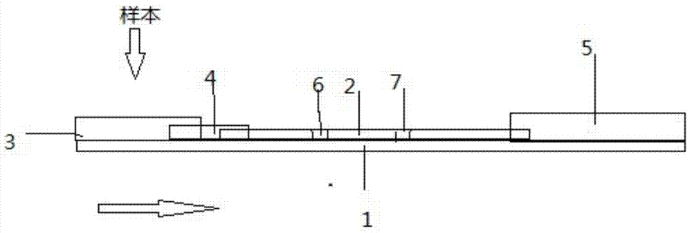
[0084] It consists of rigid polyvinyl chloride backing plate 1, nitrocellulose membrane 2, colloidal gold binding pad 4, sample pad 3, water-absorbing pad 5, detection line 6, and quality control line 7. It is characterized in that: nitrocellulose membrane 2 Pasted on the rigid polyvinyl chloride backing plate 1, on the left side of the nitrocellulose membrane 2, there are colloidal gold bonding pads 4, sample pads 3, and water-absorbing pads 5, which are arranged on the nitrocellulose membrane 2. The upper right position. Colloidal gold binding pad 4 is made of glass cellulose membrane, what sprayed on this colloidal gold binding pad 4 is the monoclonal antibody 1-E11 (hybridoma cell line 1-E11 of the anti-Duck Tembusu virus of colloidal gold labeling, preservation No. CCTCC NO:C2014219 secretion), the nitrocellulose membrane 2 is the detection membrane of the test strip, the upper left is the det...

Embodiment 3

[0086] A preparation method for detecting duck Tembusu virus immune colloidal gold test strip, the steps are:

[0087] (1) According to the method of Example 1, duck Tembusu virus monoclonal antibody was prepared.

[0088] (2) Prepare colloidal gold by reacting trisodium citrate with chloroauric acid: put 125mL water for injection into the silicified bottle, then add 1.2mL 1% (w / v) chloroauric acid, and heat in a microwave oven for 4 minutes; Quickly add 1.8 mL of 1.05% (w / v) trisodium citrate aqueous solution at one time, heat on medium heat for 5 minutes, and cool naturally at room temperature to obtain a colloidal gold solution. The appearance of the prepared colloidal gold is clear and transparent wine red, the spectrum at 450-600nm is scanned by a spectrophotometer, the maximum absorption wavelength is 520nm, and the particle size is uniform when observed by an electron microscope.

[0089] (3) Duck Tembusu virus monoclonal antibody colloidal gold labeling:

[0090] (a)...


PUM


Abstract

The invention provides a monoclonal antibody for a duck tembusu virus and application. A hybridoma cell strain 1-E11 with the collection number CCTCCNO being C2014219 and a hybridoma cell strain 4-C3 with the collection number CCTCCNO being C2014220 of the monoclonal antibody resisting the duck tembusu virus are obtained by using a purified duck-tembusu-virus immunized Balb / c mouse; a colloidal-gold test strip is prepared by utilizing the monoclonal antibody secreted by the cell strains, the 1-E11 is adopted as a labeled monoclonal antibody, 4-C3 is used as a coated monoclonal antibody, and the obtained test strip has the characteristics of fast and sensitive detection and good specificity. A result is clear and the judgment is easy; the carry and the use are convenient, and the detection cost for disease is saved.

Description

technical field [0001] The invention relates to the technical field of animal virology and immunology detection, in particular to a duck Tembusu virus monoclonal antibody and its application. Background technique [0002] Duck tembusu virus disease is a new infectious disease caused by Duck tembus virus (DTMUV), a genus of Flaviviridae, characterized by duck death and laying duck egg production decline. Other viruses of the Flavivirus genus include Japanese encephalitis virus, West Nile virus, and dengue virus. [0003] Since 2010, infectious diseases characterized by feed reduction, egg production decline and certain death rate have occurred in most breeding duck and laying duck breeding areas in my country. The large-scale outbreak of the disease first occurred from April to June 2010, and the incidences were mainly concentrated in Fujian, Zhejiang, Anhui and other places where breeding ducks and laying ducks were densely raised, and then quickly spread to Henan, Shandong...

Claims


Application Information

Patent Timeline
Patent Type & Authority Applications(China)
IPC IPC(8): G01N33/577
CPCC07K16/10G01N33/558G01N33/56983G01N2333/183
Inventor 金梅林陈伦勇
Owner HUAZHONG AGRI UNIV
Why Eureka
Try Eureka