Method for manufacturing medicine balloons

A technology of balloons and drugs, applied in catheters, coatings, etc., to achieve high drug content, maintain mechanical properties, and increase the effect of contact area

Active Publication Date: 2015-09-23
ZHEJIANG BARTY MEDICAL TECH CO LTD
View PDF4 Cites 10 Cited by
  • Summary
  • Abstract
  • Description
  • Claims
  • Application Information

AI Technical Summary

Problems solved by technology

[0002] The emergence of drug-coated stents is a milestone in the history of coronary intervention, because it reduces in-stent restenosis. However, drug-coated stents still have a stenosis rate of 5% to 10%. For special lesions, this stenosis rate is even higher. high

Method used


Image

  • Method for manufacturing medicine balloons
  • Method for manufacturing medicine balloons
  • Method for manufacturing medicine balloons

Examples


Embodiment 1

[0041] (1) Preparation of drug solution: Add 0.15 g of paclitaxel and 0.3 g of PVP into 10 mL of ethanol, mix well, and heat at 40° C. until paclitaxel and PVP are completely dissolved.

[0042] (2) Particle coating preparation: take a balloon with a size of 4.0mm*60mm, and use an ultrasonic spraying process to spray a sodium chloride solution with a concentration of 0.5 mg / mL on the surface of the balloon, and obtain a 1 μm thick particle coating after drying.

[0043] (3) Balloon drug coating: the balloon with particle coating is immersed in the drug solution in step (1), and the temperature of the drug solution is lowered to -20° C., and kept for 1 hour.

[0044] (4) The balloon was slowly taken out, and the balloon was left to dry at 40° C. for 30 minutes to obtain a drug balloon with a drug coating thickness of 10 μm.

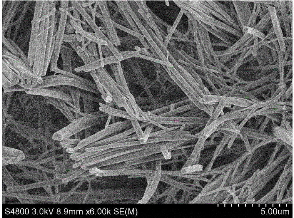
[0045] figure 1 It is the SEM picture of the surface of the drug balloon prepared in Example 1. It can be seen from the picture that the surface of the d...

Embodiment 2

[0047] (1) Preparation of drug solution: Add 0.5 g of paclitaxel and 0.3 g of PEG into 10 mL of methanol, mix well, and heat at 60° C. until paclitaxel and PEG are completely dissolved.

[0048](2) Balloon particle coating preparation: take a balloon with a size of 3.0mm*20mm, use ultrasonic spraying process, spray calcium chloride solution with a concentration of 10mg / mL on the surface of the balloon, and obtain a 5μm thick particle coating after drying .

[0049] (3) Add 1ml of water for injection to the solution in step (1), stir well and let stand for 1 minute.

[0050] (4) Submerge the microparticle-coated balloon in the solution of step (3), and keep at room temperature for 1 hour.

[0051] (5) The above-mentioned balloon was slowly taken out, and the balloon was left to dry at room temperature for 1 hour to obtain a drug-coated balloon with a drug-coating thickness of 20 μm.

Embodiment 3

[0053] (1) Preparation of drug solution: 0.13 g of rapamycin and 0.3 g of PVA were added into 10 ml of acetonitrile, stirred to completely dissolve the drug.

[0054] (2) Balloon particle coating preparation: immerse a 4.0mm*30mm balloon in 20mg / mL urea solution, take out the balloon slowly after 10 minutes, place it at 40°C, and obtain a 30μm thick particle coating after drying ;

[0055] (3) Balloon drug coating: the balloon with microparticle coating is immersed in the drug solution in step (1), and the temperature of the drug solution is lowered to -60° C. and kept for 30 minutes.

[0056] (4) The balloon was slowly taken out, and the balloon was left to dry at room temperature for 30 minutes to obtain a drug balloon with a drug coating thickness of 15 μm.


PUM

PropertyMeasurementUnit
thicknessaaaaaaaaaa
thicknessaaaaaaaaaa
concentrationaaaaaaaaaa

Abstract

The invention provides a method for manufacturing medicine balloons. The method includes steps of (1), manufacturing particle coatings on the surfaces of balloons. (2), arranging the balloons with the particle coatings in medicine liquor, crystallizing medicine on the surfaces of the particle coatings to form medicine coatings, taking the balloons out of the medicine liquor and drying the balloons to obtain the medicine balloons. The particle coatings are made of sodium chloride or calcium chloride or urea. The method has the advantages that the particle coatings are manufactured, accordingly, the formed medicine coatings are uniform, and the medicine coatings and the surfaces of the balloons are good in affinity; the coatings rarely fall off when the medicine balloons are folded, compressed and gripped, and the medicine can be quickly transferred to the blood vessel walls after short contact and can be retained on the blood vessel walls for a long time.

Description

technical field [0001] The invention relates to the technical field of medical device preparation, in particular to a preparation method of a drug balloon. Background technique [0002] The emergence of drug-coated stents is a milestone in the history of coronary intervention because it reduces in-stent restenosis. However, drug-coated stents still have a stenosis rate of 5% to 10%. For special lesions, this stenosis rate is even higher. high. Moreover, in recent years, it has been found that there is late stent thrombosis in drug stents, which has led to the clinical demand for new medical devices. The drug-coated balloon is an interventional medical device. After the drug is delivered in the blood vessel, it is withdrawn from the blood vessel, and the drug stays in the lesion. Through the continuous release of the drug, it can inhibit the stenosis of the blood vessel. This is a very ideal new medical device. , has become a new research hotspot in the field of vascular in...

Claims


Application Information

Patent Timeline
Patent Type & Authority Applications(China)
IPC IPC(8): A61L29/16A61L29/10A61L29/08
CPCA61L29/08A61L29/10A61L29/16
Inventor 张芝芳
Owner ZHEJIANG BARTY MEDICAL TECH CO LTD
Why Eureka
Try Eureka