Method of quickly separating breast cancer primary tumor living cell

A breast cancer and living cell technology, applied in the field of cell separation, can solve the problems of fibroblast contamination, low purity of separation of breast cancer cells, inability to use clinical drug sensitivity tests, etc., and achieve the effect of broad material sources

Active Publication Date: 2015-10-14
上海兰卫医学检验所股份有限公司
View PDF0 Cites 1 Cited by
  • Summary
  • Abstract
  • Description
  • Claims
  • Application Information

AI Technical Summary

Problems solved by technology

[0025] The present invention aims at the problem that primary breast cancer cells have low separation purity and are easily contaminated by fibroblasts in the current field of biological and medical research, so that they cannot be used in clinical drug sensitivity tests, and propose a high Method for Rapid Separation of Pure Batch Live Primary Tumor Cells of Breast Cancer

Method used


Image

  • Method of quickly separating breast cancer primary tumor living cell

Examples


Embodiment 1

[0058] Embodiment 1 sample collection

[0059] All samples used in the implementation of the present invention are derived from surgical resection specimens of breast cancer patients, and all participating patients have given informed consent to the scientific research.

[0060] The detection samples used in the present invention are usually collected in a clinically acceptable manner, preferably in a manner in which nucleic acid (especially RNA) or protein is protected.

[0061] Patient information (age, gender, impact reports, course of treatment, other medical conditions, family history, etc.) was obtained from the hospital database and used to match the various samples collected. Pathological follow-up studies (such as histological analysis by hematoxylin and eosin staining, ie H&E staining) are used to clarify the disease status of a given sample and to ensure consistent grading of samples.

[0062] Fresh breast cancer specimens were obtained in the operating room and se...

Embodiment 2

[0063] Embodiment 2: tissue pretreatment

[0064] The pathologist cut out a 1cm3 tissue block from the non-necrotic area of ​​the sample to be tested and the area containing more tumor tissue. At the same time, the tissue block was subjected to frozen section and HE staining for quality control to ensure that the tumor cell content reached more than 75%. Place the tissue in 10ml of 199 culture medium containing gentamicin, and store it on ice for 24 hours for specimen transportation.

Embodiment 3

[0065] Example 3: Rapid batch separation of high-purity primary breast cancer cells

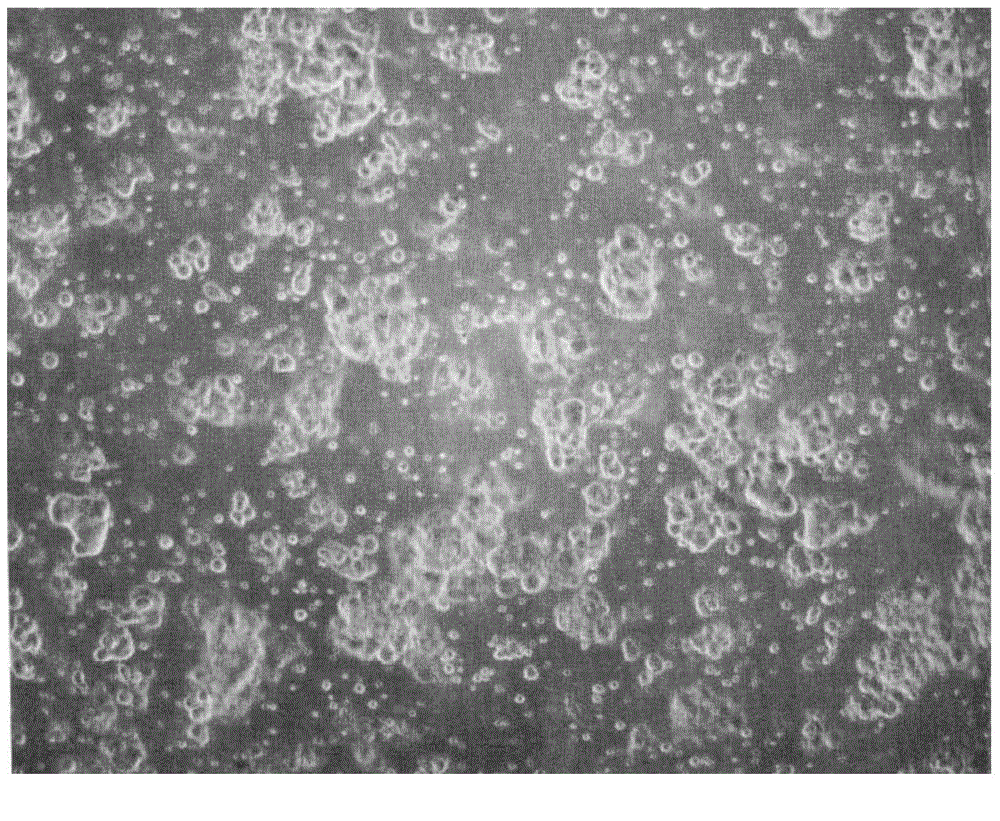
[0066] 1cm 3 Large and small breast cancer tissue blocks cut into 1mm 3 For tissue fragments of different sizes, wash the tissue twice with 10ml of 199 culture medium containing gentamicin, and repeatedly pipette for 2 minutes, transfer 10ml of 199 culture medium containing tissue to a 15ml centrifuge tube and let it stand for 1 minute, carefully Remove the supernatant to another clean 15ml centrifuge tube, and avoid aspirating the tissue pieces at the bottom, centrifuge the supernatant obtained above at a speed of 1000RPM for 5 minutes, discard the supernatant carefully, and use 10ml of Ham'sF-12 Resuspend the cell pellet in the culture medium, in which Ham'sF-12 culture medium contains 5% FBS, 1 μg / ml insulin and 1 μg / ml hydrocortisone, move the obtained cells to a culture dish, at 37oC, 9% CO 2 cultured in an environment, observe the obtained cells with a microscope, count and take pi...


PUM

PropertyMeasurementUnit
purityaaaaaaaaaa

Abstract

The invention belongs to the technical field of cell separation, and relates to a method of quickly separating breast cancer primary tumor living cells in batches at a high purity. The method includes the steps of: (1) obtaining an isolated breast cancer fresh tissue sample; (2) performing pretreatment and quality control of the tissue sample; and (3) quickly separating the breast cancer primary tumor living cells in batches at a high purity, so that the high-purity breast cancer primary tumor living cells are separated in batches quickly. According to the invention, living breast cancer cells are separated from breast cancer tissue blocks with the purity of the tumor cells being higher than 90%. The separated breast cancer primary tumor living cells can be used in drug sensitive tests. The method overcomes the defects that the breast cancer primary tumor living cells are low in separation purity and are very liable to be polluted by fibroblast and can provide reliable research object for clinical drug sensitive tests. The invention provides the new method and wider material sources for database foundation of DNA, RNA and protein of primary tumor living cells and tumor in-vivo researching.

Description

technical field [0001] The invention belongs to the technical field of cell separation, and relates to a method for separating live primary tumor cells of breast cancer, in particular to a method for rapidly separating high-purity batch live primary tumor cells of breast cancer. The isolated living primary tumor cells of breast cancer can be used for drug sensitivity test. Background technique [0002] According to reports, the vast majority of malignant tumor patients are treated with chemical drugs after surgery at home and abroad. Studies have shown that chemotherapy is only effective for about 25% of cancer patients, and for the remaining 75% of patients who are ineffective, it is undoubtedly suffering and aggravating the economic and mental pressure on the family and society. Studies have shown that stimulation of chemotherapy drugs to those breast cancer cells that are not sensitive to chemotherapy drugs will promote EMT and metastasis of tumor cells. It can be seen t...

Claims


Application Information

Patent Timeline
Patent Type & Authority Applications(China)
IPC IPC(8): C12N5/09C12Q1/02
Inventor 朱虹光王漱阳陈琦王维
Owner 上海兰卫医学检验所股份有限公司
Why Eureka
Try Eureka