Method for observing morphology of ultrafine powder sample by using scanning electron microscope

An electron microscope, ultra-fine powder technology, applied in the direction of analyzing materials, instruments, etc., to achieve good dispersion, good effect, and good detection selectivity.

Inactive Publication Date: 2015-10-21
OCEAN UNIV OF CHINA
View PDF4 Cites 13 Cited by
  • Summary
  • Abstract
  • Description
  • Claims
  • Application Information

AI Technical Summary

Problems solved by technology

[0004] The purpose of the present invention is to provide an analytical method for observing the morphology of ultrafine powdered samples with a scanning electron microscope, which is simple and easy to implement, aiming at the deficiencies of the existing scanning electron microscope for observing the morphology of ultrafine powder samples. It can realize the rapid and selective detection of ultrafine powder, and the detection process does not require other reagents other than the sample, etc.

Method used


Image

  • Method for observing morphology of ultrafine powder sample by using scanning electron microscope
  • Method for observing morphology of ultrafine powder sample by using scanning electron microscope
  • Method for observing morphology of ultrafine powder sample by using scanning electron microscope

Examples


Embodiment 1

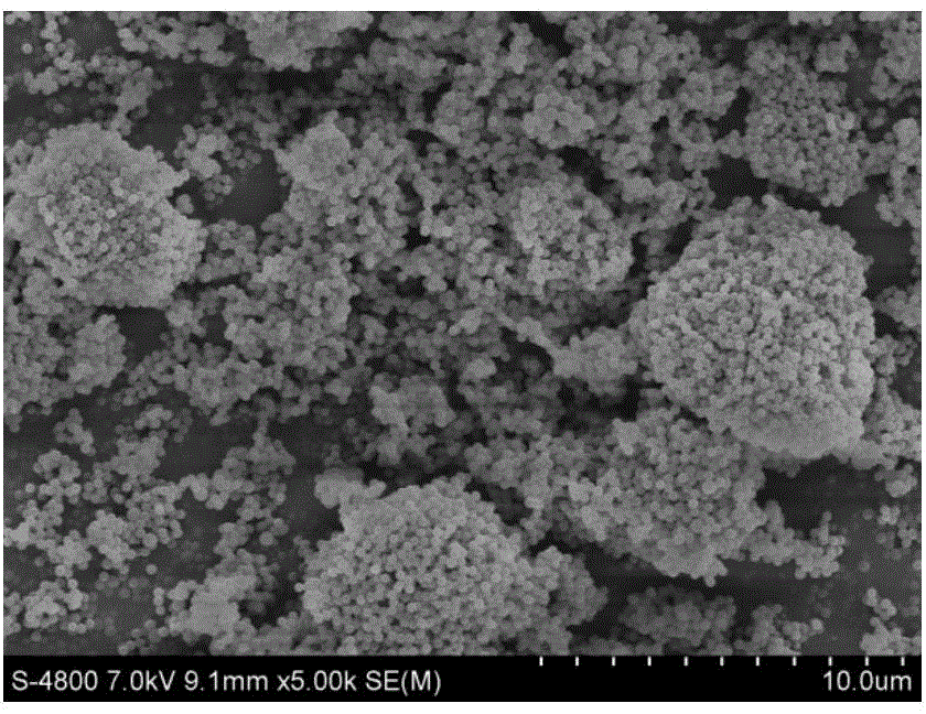
[0029] The specific steps of the analysis method of the superfine powder sample PVDF morphology observation that the present embodiment provides are as follows:

[0030] 1) Evenly spread the superfine powder sample PVDF20mg~40mg on the weighing paper;

[0031] 2) After rubbing two pieces of plastic film (area 10cm×10cm, thickness 50-80μm) to generate static electricity, place the charged side of one of the plastic films horizontally above the powder, about 2cm away from the PVDF powder, and use electrostatic force to absorb a part powder on plastic film; on film;

[0032] 3) Cut a piece of double-sided conductive tape, and use one side of the tape to glue the electrostatically adsorbed powder on the film;

[0033] 4) Use compressed air to fully purge the PVDF powder bonded to the double-sided conductive tape, so that only a small amount of powder with strong adhesion to the conductive tape remains;

[0034] 5) Peel off the protective layer on the other side of the double-sid...

Embodiment 2

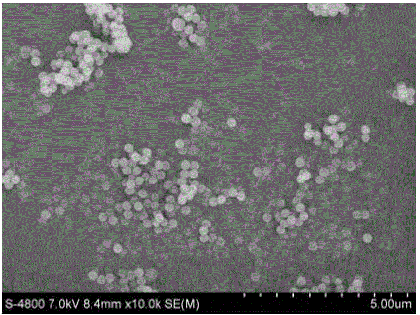
[0038] The specific steps of the analysis method for the observation of the nanoscale synthetic silica morphology of the ultrafine powder sample provided in this example are as follows:

[0039] 1) Evenly spread 20mg-40mg of superfine powder sample silicon dioxide on the weighing paper;

[0040] 2) After rubbing two pieces of plastic film (10cmX10cm) to generate static electricity, place the charged side of one of the plastic films horizontally above the powder, about 2cm away from the silica powder, and use electrostatic force to absorb a part of the powder on the film;

[0041] 3) Cut a piece of double-sided conductive tape, and use one side of the tape to glue the electrostatically adsorbed powder on the film;

[0042] 4) Use compressed air to fully purge the silica powder bonded to the double-sided conductive tape, so that only a small amount of powder with strong adhesion to the conductive tape remains;

[0043] 5) Peel off the protective layer on the other side of the d...

Embodiment 3

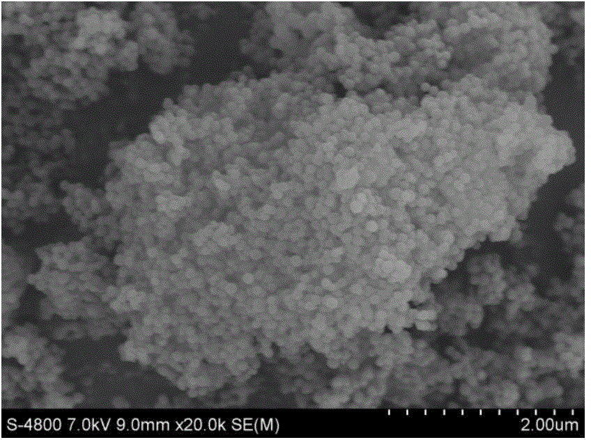
[0047] The specific steps of the analysis method for the observation of the carbon nanotube powder morphology provided by the present embodiment are as follows:

[0048] 1) Evenly spread 20mg to 40mg of carbon nanotube powder on the weighing paper;

[0049] 2) After rubbing two pieces of plastic film (10cmX10cm) to generate static electricity, place the charged side of one of the plastic films horizontally above the powder, about 2cm away from the silica powder, and use electrostatic force to absorb a part of the powder on the film;

[0050] 3) Cut a piece of double-sided conductive tape, and use one side of the tape to glue the electrostatically adsorbed powder on the film;

[0051] 4) Use compressed air to fully purge the silica powder bonded to the double-sided conductive tape, so that only a small amount of powder with strong adhesion to the conductive tape remains;

[0052] 5) Peel off the protective layer on the other side of the double-sided conductive tape, and stick ...


PUM

PropertyMeasurementUnit
particle sizeaaaaaaaaaa
sizeaaaaaaaaaa

Abstract

The objective of the invention is to provide an analytical method for observing the morphology of an ultrafine powder sample by using a scanning electron microscope. The method provided by the invention comprises the following steps: 1) adsorbing the ultrafine powder sample by using an electrostatically charged smooth surface of an adsorbate; 2) bonding powder adsorbed onto the adsorbate, i.e., a film, with one side of a double-sided conducting adhesive tape; 3) adhering the other side of the double-sided conducting adhesive tape onto a sample table of the scanning electron microscope; 4) carrying out gold plating on the powder adhered on the double-sided conducting adhesive tape; and 5) carrying out observation with the scanning electron microscope. With the method, a part of powder with a uniform particle size in the ultrafine powder sample can be obtained, the powder adsorbed under the action of electrostatic attraction is fine particles in the ultrafine powder sample, and only particles with similar particle sizes can be adsorbed at the same time, so a picture with uniform granularity can be obtained in observation with the scanning electron microscope.

Description

technical field [0001] The invention belongs to the technical field of electron microscope detection, and in particular relates to a method for observing the morphology of an ultrafine powder sample with a scanning electron microscope. Background technique [0002] As an advanced analysis and testing instrument, the scanning electron microscope is mainly used for microscopic morphology observation and preliminary determination of components. It has the characteristics of simple sample preparation, adjustable magnification, wide range, and large depth of field. It has been widely used in materials It is a basic instrument widely used in modern scientific research in the fields of fracture analysis, micro-region composition analysis, various coating surface topography analysis, layer thickness measurement, microstructure topography and nano-material analysis. [0003] Ultrafine powder refers to the powder whose particle size is less than 10 μm. It was Ryoji Ueda of Japan who ...

Claims


Application Information

Patent Timeline
Patent Type & Authority Applications(China)
IPC IPC(8): G01N15/00
Inventor 毛丽莉孟春霞
Owner OCEAN UNIV OF CHINA
Why Eureka
Try Eureka