

DC cell culture reagent and culture method thereof
A cell culture and culture method technology, applied in animal cells, vertebrate cells, blood/immune system cells, etc., can solve the problem that the human immune system has little anti-tumor effect, cannot achieve antigen clearance function, and lacks activation of aggregated T cells. Cell and other issues, to achieve good cell viability and purity, good cell viability, and the effect of promoting proliferation
- Summary
- Abstract
- Description
- Claims
- Application Information
AI Technical Summary
Problems solved by technology
Method used
Image

Examples
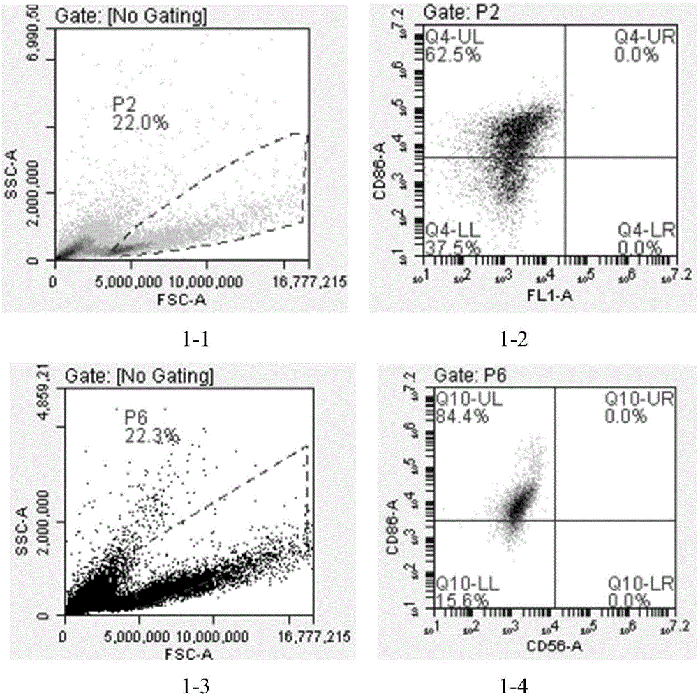
Embodiment 1D
[0063] Example 1 DC-CIK cell co-culture example
[0064] (1) Mononuclear cell isolation
[0065] 1. Peripheral blood or umbilical cord blood 40mL, centrifuge at 500-800g for 10 minutes, absorb the upper plasma, inactivate at 56°C for 30min, centrifuge at 2000-3000g for 5min, supernatant plasma at 2-8°C for later use;
[0066] 2. For the lower layer of blood, add 2 times the volume of normal saline to resuspend. According to the diluted blood: Ficoll separation solution ratio of 2:1, slowly add the diluted blood to the upper layer of Ficoll separation solution to make the layer clear;
[0067] 3. Centrifuge at 500-700g for 15-30min, and absorb the middle buffy coat layer, which is mononuclear cells.
[0068] (2) DC cell isolation and culture
[0069] 1. Dilute the obtained mononuclear cells with normal saline, centrifuge at 200-400g for 5min, and wash twice;
[0070] 2. Add serum-free medium (containing 5% autologous plasma) to resuspend the cells, press 2-8×10 6 / mL inocul...
Embodiment 2D
[0073] Example 2 DC-CIK cell co-culture example
[0074] (1) Mononuclear cell isolation
[0075] 1. Peripheral blood or umbilical cord blood 40mL, centrifuge at 500-800g for 10 minutes, absorb the upper plasma, inactivate at 56°C for 30min, centrifuge at 2000-3000g for 5min, supernatant plasma at 2-8°C for later use;
[0076] 2. For the lower layer of blood, add 2 times the volume of normal saline to resuspend. According to the ratio of diluted blood: Ficoll separation solution 2:1, slowly add the diluted blood to the upper layer of Ficoll separation solution to make the layer clear;
[0077] 3. Centrifuge at 500-700g for 15-30min, and absorb the middle buffy coat layer, which is mononuclear cells.
[0078] (2) DC cell isolation and culture
[0079] 1. Dilute the obtained mononuclear cells with normal saline, centrifuge at 200-400g for 5min, and wash twice;
[0080] 2. Add serum-free medium (containing 15% autologous plasma) to resuspend the cells, press 2-8×10 6 / mL inocu...
Embodiment 3
[0083] Embodiment 3DC-CIK cell co-culture example
[0084] (1) Mononuclear cell isolation
[0085] 1. Peripheral blood or umbilical cord blood 40mL, centrifuge at 500-800g for 10 minutes, absorb the upper plasma, inactivate at 56°C for 30min, centrifuge at 2000-3000g for 5min, supernatant plasma at 2-8°C for later use;
[0086] 2. For the lower layer of blood, add 2 times the volume of normal saline to resuspend. According to the ratio of diluted blood: Ficoll separation solution 2:1, slowly add the diluted blood to the upper layer of Ficoll separation solution to make the layer clear;
[0087] 3. Centrifuge at 500-700g for 15-30min, and absorb the middle buffy coat layer, which is mononuclear cells.
[0088] (2) DC cell isolation and culture
[0089] 1. Dilute the obtained mononuclear cells with normal saline, centrifuge at 200-400g for 5min, and wash twice;
[0090] 2. Add serum-free medium (containing 10% autologous plasma) to resuspend the cells, press 2-8×10 6 / mL ino...
PUM
 Abstract
Description
Claims
Application Information