Special culture medium and culture method for long-term maintenance, propagation, and subcultring of human hepatocyte
A technology of subculture and culture medium, applied in artificial cell constructs, animal cells, vertebrate cells, etc., can solve the problems that liver cells cannot be used as a qualified cell model for liver disease research, complicated operations, and impossible to source liver cells.
- Summary
- Abstract
- Description
- Claims
- Application Information
AI Technical Summary
Problems solved by technology
Method used
Image
Examples
Embodiment 1
[0092] Embodiment 1, preparation human hepatocyte culture medium
[0093] 1. Hepatocyte basal medium was prepared according to conventional methods, that is, the ingredients in Table 4 were added to DMEM / F12 (basic cell culture medium) (percentages are in v / v):
[0094] Table 4
[0095] N2
2.5%
B27
5%
Non-essential amino acids (Non-AA)
1%
Sodium pyruvate
1%
Penicillin and streptomycin mixture (100x)
1%
[0096] 2. On the basis of the basic medium for hepatocytes above, add the components with final concentrations as shown in Table 5 to obtain hepatocyte culture medium I-1.1 / 2, I-2.1 / 2 and hepatocyte culture medium I-3.1 respectively / 2 / 3 / 4.
[0097] table 5
[0098]
Embodiment 2
[0099] Example 2, long-term proliferation, subculture and maintenance of human hepatocytes
[0100]1. Hepatocyte Culture Medium Ⅰ-1.1, Hepatocyte Culture Medium Ⅰ-1.2 Culture of primary human hepatocytes
[0101] (1) Primary hepatocytes (separated from excised human liver tissue) were purchased from Lifetechnology Company.
[0102] (2) Prime the culture plate with matrigel (1:30) for 1 hour, then suspend the primary hepatocytes in the adherent medium (basic medium for hepatocytes supplemented with 10% serum), plate and store at 37°C Incubate for 1 hour;
[0103] (3) The culture medium in (2) was discarded, and the hepatocytes were cultured in hepatocyte medium I-1.1 and I-1.2 respectively.
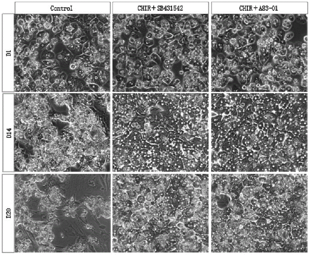
[0104] Using hepatocyte culture medium Ⅰ-1.1 and Ⅰ-1.2, when cultured for 1, 14 and 20 days, the cell morphology was taken (the cells cultured without adding GSK3β inhibitor and TGFβ inhibitor were used as control), and the morphology diagram was as follows: figure 1 .
[0105] Depend ...
Embodiment 3
[0124] Embodiment 3, identification of liver cell characteristics
[0125] (1) Immunostaining
[0126] The immunostaining method is:
[0127] 1. Discard the cell culture medium, rinse with PBS once,
[0128] After fixing with 2.4% paraformaldehyde for 10 minutes, rinse with PBS for 5 minutes x 3 times,
[0129] 3. 10% goat serum blocking: room temperature, 60 minutes,
[0130] 4.0.2% Triton: 25-30min,
[0131] 5. Primary antibody (rabbit anti-ALB antibody, mouse anti-CYP3A antibody or rabbit anti-Asgpr antibody) at room temperature for 1 hour
[0132] or overnight at 4°C,
[0133] 6. Rinse with PBS for 5 minutes x 3 times,
[0134] 7. Secondary antibody (Cy3-labeled goat anti-rabbit antibody, FITC-labeled goat anti-mouse or goat anti-rabbit antibody) at room temperature
[0135] 45-60min,
[0136] 8. Wash with PBS, 5min×3 times,
[0137] 9. Seal the slides and take pictures with a fluorescence microscope.
[0138] The cells cultured in vitro for 84 days were immunost...
PUM
 Abstract
Description
Claims
Application Information
 - R&D
- Intellectual Property
- Life Sciences
- Materials
- Tech Scout
- Unparalleled Data Quality
- Higher Quality Content
- 60% Fewer Hallucinations
Browse by: Latest US Patents, China's latest patents, Technical Efficacy Thesaurus, Application Domain, Technology Topic, Popular Technical Reports.
© 2025 PatSnap. All rights reserved.Legal|Privacy policy|Modern Slavery Act Transparency Statement|Sitemap|About US| Contact US: help@patsnap.com


