Drug-delivery endovascular stent and method for treating restenosis

a technology of endovascular stent and drug delivery, which is applied in the field of endovascular drug delivery stent and a method for treating restenosis, can solve the problems of reducing or eliminating the beneficial effect of angioplasty/stenting procedure, blood clots may also form inside the newly implanted stent, and normal damage to endothelial cells which line the inside surface of the vessel lumen

Inactive Publication Date: 2005-09-06
BIOSENSORS INT GROUP
View PDF23 Cites 321 Cited by
  • Summary
  • Abstract
  • Description
  • Claims
  • Application Information

AI Technical Summary

Benefits of technology

[0011]The invention includes, in one aspect, an endovascular stent for placement at a vascular injury site, for inhibiting restenosis at the site. The stent is constructed of a structural member or body formed of one or more filaments and carried on the stent-body filament(s), a bioerodable drug-release coating having a thickness of between 3-15 microns, and composed of (i) 20 and 60 weight percent poly-dl-lactide polymer substrate and (ii) 40-80 weight percent of an anti-restenosis compound. A polymer undercoat having a thickness of between 1-5 microns and disposed between the stent-body filaments and the coating helps to stabilize the coating on the stent filaments. The stent is expandable from a contracted condition in which the stent can be delivered to a vascular injury site via catheter, and an expanded condition in which the stent coating can be placed in contact with the vessel at the injury site. The stent coating is effective to release a restenosis-inhibiting amount of the compound over a period of at least 4 weeks after the stent is placed at the vascular injury site.
[0016]The stent is expandable from a contracted condition in which the stent can be delivered to a vascular injury site via catheter, and an expanded condition in which the stent coating can be placed in contact with the vessel at the injury site. The coating is effective to release the restenosis-inhibiting amount of the compound over a period of at least 4 weeks after the stent is placed at the vascular injury site.

Problems solved by technology

During the healing process, inflammation caused by angioplasty and stent implant injury often causes smooth muscle cell proliferation and regrowth inside the stent, thus partially closing the flow channel, and thereby reducing or eliminating the beneficial effect of the angioplasty / stenting procedure.
Blood clots may also form inside of the newly implanted stent due to the thrombotic nature of the stent surfaces, even when biocompatible materials are used to form the stent.
A heparinized surface is known to interfere with the blood-clotting cascade in humans, preventing attachment of platelets (a precursor to thrombin) on the stent surface.
Finally, endothelial cells which line the inside surface of the vessel lumen are normally damaged by the process of angioplasty and / or stenting.

Method used


Image

  • Drug-delivery endovascular stent and method for treating restenosis
  • Drug-delivery endovascular stent and method for treating restenosis
  • Drug-delivery endovascular stent and method for treating restenosis

Examples


example 1

Preparation of Everolimus and Derivatives Thereof

Step A. Synthesis of 2-(t-butyldimethylsilyl)oxyethanol(TBS glycol).

[0101]154 ml of dry THF and 1.88 g NaH are stirred under in a nitrogen atmosphere in a 500 ml round bottom flask condenser. 4.4 mL dry ethylene glycol are added into the flask, resulting in a large precipitate after 45 minutes of stirring. 11.8 g tert-butyldimethylsilyl chloride is added to the flask and vigorous stirring is continued for 45 minutes. The resulting mixture is poured into 950 mL ethylether. The ether is washed with 420 mL brine and solution is dried with sodium sulfate. The product is concentrated by evaporation of the ether in vacuo and purified by flash chromatography using a 27×5.75 cm column charged with silica gel using a hexanes / Et2O (75:25v / v) solvent system. The product is stored at 0° C.

Step B. Synthesis of 2-(t-butyldimethylsilyl)oxyethyl Triflate (TBS Glycol Trif)

[0102]4.22 g TBS glycol and 5.2 g 2,6-lutidine are combined in a double-necked 1...

example 2

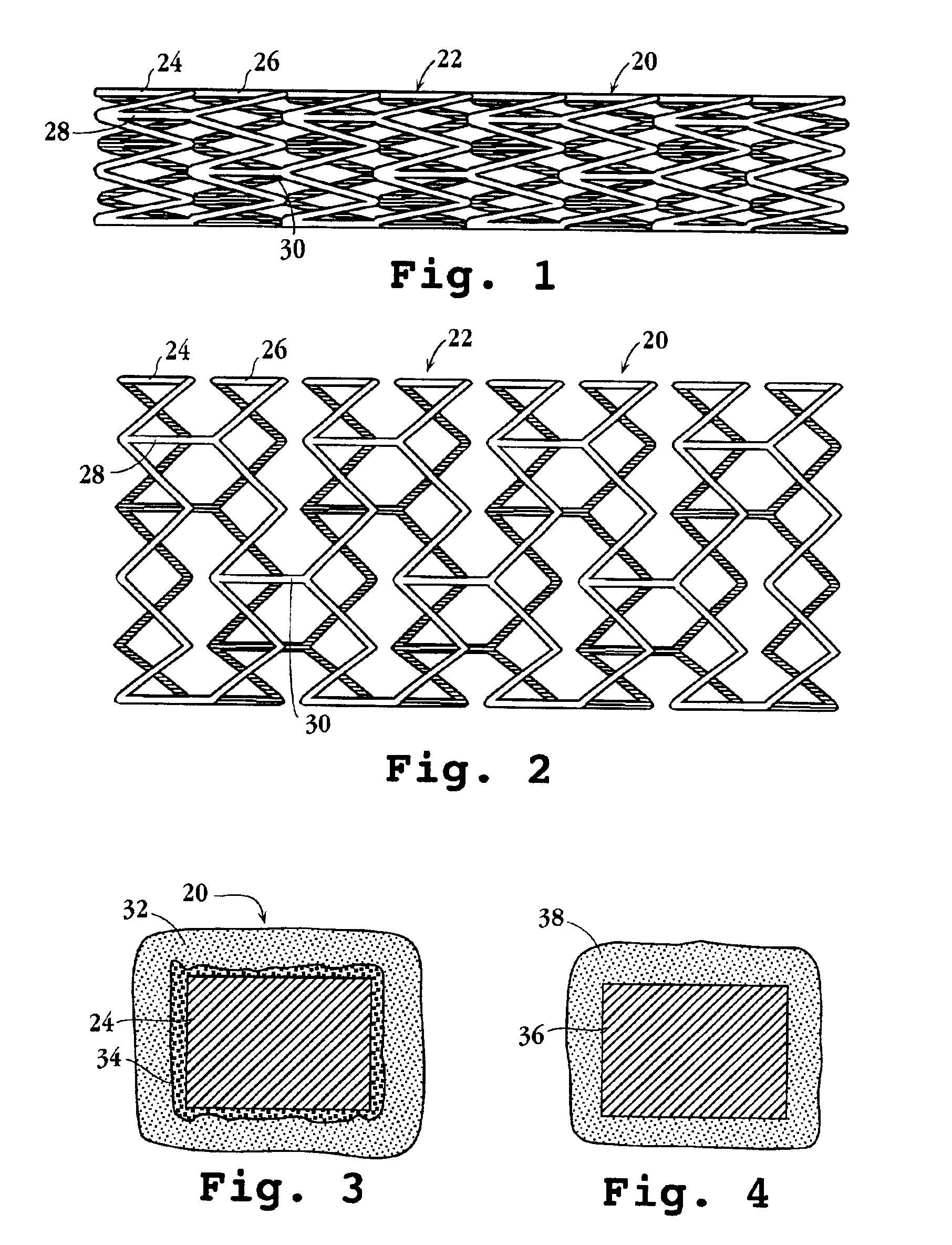
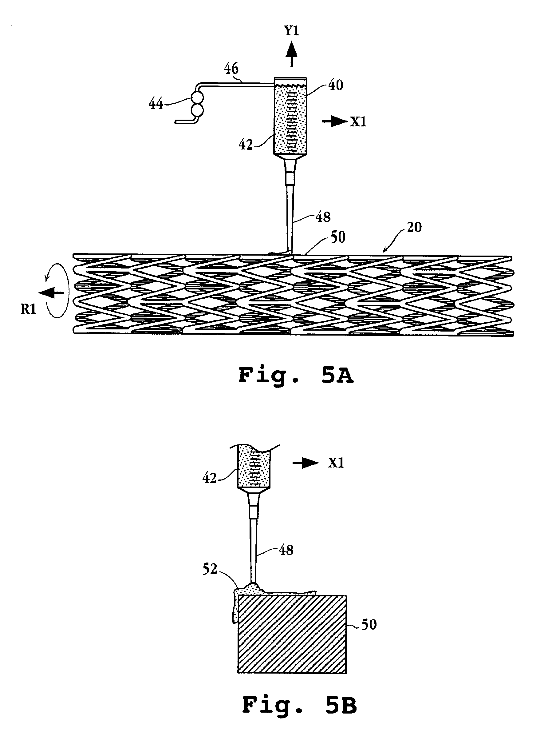
Preparation of Stent Containing Everolimus in a poly-dl-lactide Coating

[0105]100 mg poly (dl-lactide) was dissolved into 2 mL acetone at room temperature. 5 mg everolimus was placed in a vial and 400 μL lactide solution added. A microprocessor-controlled syringe pump was used to precision dispense 10 μL of the drug containing lactide solution to the stent strut top surfaces. Evaporation of the solvent resulted in a uniform, drug containing single polymer layer on the stent.

[0106]A 15 μL volume was used in a similar manner to coat the stent top and side strut surfaces, resulting in a single layer coating on the stent strut top and sides.

example 3

In Vitro Drug Release from Stent Containing Everolimus in a poly-dl-lactide Coating

[0107]In vitro drug release was conducted by placing the coated stents into 2 mL pH 7.4 phosphate buffered saline solution containing 25% ETOH, and preserved with 0.05% (w / v) sodium azide and maintained at 37° C. Sampling was periodically conducted by withdrawing the total buffer volume for drug measurement while replacing solution with a similar volume of fresh buffer (infinite sink). FIG. 7 illustrates drug release from two similar stents coated with a single polymer layer microdispensed in this manner.


PUM

PropertyMeasurementUnit
thicknessaaaaaaaaaa
thicknessaaaaaaaaaa
thicknessaaaaaaaaaa

Abstract

An intravascular stent and method for inhibiting restenosis, following vascular injury, is disclosed. The stent has an expandable, linked-filament body and a drug-release coating formed on the stent-body filaments, for contacting the vessel injury site when the stent is placed in-situ in an expanded condition. The coating releases, for a period of at least 4 weeks, a restenosis-inhibiting amount of a monocyclic triene immunosuppressive compound having an alkyl group substituent at carbon position 40 in the compound. The stent, when used to treat a vascular injury, gives good protection against clinical restenosis, even when the extent of vascular injury involves vessel overstretching by more than 30% diameter. Also disclosed is a stent having a drug-release coating composed of (i) 10 and 60 weight percent poly-dl-lactide polymer substrate and (ii) 40-90 weight percent of an anti-restenosis compound, and a polymer undercoat having a thickness of between 1-5 microns.

Description

FIELD OF THE INVENTION[0001]The present invention relates to an endovascular drug-delivery stent and to a method for treating restenosis.BACKGROUND OF THE INVENTION[0002]A stent is a type of endovascular implant, usually generally tubular in shape, typically having a lattice, connected-wire tubular construction which is expandable to be permanently inserted into a blood vessel to provide mechanical support to the vessel and to maintain or re-establish a flow channel during or following angioplasty. The support structure of the stent is designed to prevent early collapse of a vessel that has been weakened and damaged by angioplasty. Insertion of stents has been shown to prevent negative remodeling and spasm of the vessel while healing of the damaged vessel wall proceeds over a period of months.[0003]During the healing process, inflammation caused by angioplasty and stent implant injury often causes smooth muscle cell proliferation and regrowth inside the stent, thus partially closing...

Claims


Application Information

Patent Timeline
Patent Type & Authority Patents(United States)
IPC IPC(8): A61F2/06A61K9/00A61L31/08A61L31/10A61L31/14A61L31/16A61F2/00A61K9/10A61F2/90A61K9/06A61K9/20A61K9/70A61K31/4353A61K31/436A61K47/34A61L31/00A61P9/10A61P9/14A61P17/02A61P29/00A61P35/00C07D498/18
CPCA61F2/90A61L31/06A61F2/915A61K9/0024A61L31/10A61L31/16A61F2/91A61L31/022C08L29/04C08L67/04A61F2002/072A61F2002/91533A61F2002/91575A61F2240/001A61F2250/0067A61L2300/416A61L2300/602A61L2300/606A61F2230/0054A61L2420/08A61P17/02A61P29/00A61P35/00A61P9/10A61P9/14
Inventor SHULZE, JOHN E.BETTS, RONALD E.SAVAGE, DOUGLAS R.
Owner BIOSENSORS INT GROUP
Why Eureka
Try Eureka