Method for extracting mycobacterium tuberculosis DNA from mouse tissue

A technology of Mycobacterium tuberculosis and extraction method, which is applied in the field of DNA extraction, can solve the problems of low DNA extraction rate and cumbersome operation, and achieve the effect of simple operation, high purity and concentration, and high extraction rate

Inactive Publication Date: 2016-01-06
KUNMING MEDICAL UNIVERSITY
View PDF1 Cites 1 Cited by
  • Summary
  • Abstract
  • Description
  • Claims
  • Application Information

AI Technical Summary

Problems solved by technology

The xylene and acetone extraction methods for tissue paraffin slices are cumbersome to operate, and the extraction rate of DNA is low; the kit extraction method: the DNA extraction kit purchased on the market can be used to extract DNA after tissue lysis and cell lysis, although this The method is easy to operate, but the extraction rate of DNA is low

Method used


Image

  • Method for extracting mycobacterium tuberculosis DNA from mouse tissue
  • Method for extracting mycobacterium tuberculosis DNA from mouse tissue
  • Method for extracting mycobacterium tuberculosis DNA from mouse tissue

Examples


Embodiment 1

[0025] Mouse lung tissue (80mg)

[0026] ↓ Homogenization, 10000rpm, 1min

[0027] Discard the supernatant, add 200 μl of GA (tissue lysate) and 20 μl of proteinase K solution to the pellet

[0028] ↓56°C water bath for 3.0h to obtain cell suspension

[0029] Add 200 μl of TES (the pH of the cell lysate is 8, and the concentration of lysozyme is 20 mg / ml) and

[0030] 5% (W / V) SDS (precipitating agent) 120μl

[0031] ↓36℃ water bath for 40 minutes, boiling water bath for 60 minutes, place on ice for 5 minutes,

[0032] get a suspension

[0033] Add 500μl PH4.8 NaAc (sodium acetate) and 100μl 0.05mol / LG (glucose)

[0034] ↓Invert and mix well, place on ice for 15 minutes and divide into two tubes

[0035] Add 600 μl phenol / chloroform / isoamyl alcohol (25:24:1) to each tube

[0036] ↓Inverted and mixed, 12000rpm, 10min

[0037] Transfer the upper aqueous phase to a new centrifuge tube

[0038] ↓

[0039] Add an equal volume of chloroform / isoamyl alcohol (24:1)

[0040]...

Embodiment 2

[0047] Mouse lung tissue (80mg)

[0048] ↓ Homogenization, 10000rpm, 1min

[0049] Discard the supernatant, add 200 μl of GA (tissue lysate) and 20 μl of proteinase K solution to the pellet

[0050] ↓56°C water bath for 12 hours to obtain cell suspension

[0051] Add 200 μl of TES (the pH of the cell lysate is 8, and the concentration of lysozyme is 20 mg / ml) and

[0052] 5% (W / V) SDS (precipitating agent) 120μl

[0053] ↓37℃ water bath for 40 minutes, boiling water bath for 60 minutes, and place on ice for 7 minutes to obtain a suspension

[0054] Add 500μl PH4.8 NaAc (sodium acetate) and 100μl 0.05mol / L (glucose)

[0055] ↓Invert and mix well, place on ice for 20min and divide into two tubes

[0056] Add 600 μl phenol / chloroform / isoamyl alcohol (25:24:1) to each tube

[0057] ↓Inverted and mixed, 12000rpm, 10min

[0058] Transfer the upper aqueous phase to a new centrifuge tube

[0059] ↓

[0060] Add an equal volume of chloroform / isoamyl alcohol (24:1)

[0061] ↓1...


PUM


Abstract

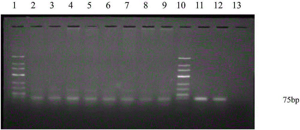
The invention discloses a method for extracting mycobacterium tuberculosis DNA from a mouse tissue. The method for extracting the mycobacterium tuberculosis DNA from the mouse tissue comprises the following steps: (1) homogenising a mouse lung tissue, centrifuging, then abandoning a supernatant, adding a GA buffer solution and a proteinase K solution, and then carrying out water bath for implementing histolysis, so as to obtain cell suspension; (2) adding a TES buffer solution (containing lysozyme) and an SDS solution into the cell suspension, putting the cell suspension in a boiling water bath for 60 minutes, and placing on ice for obtaining suspension; and (3) adding a sodium acetate solution and a glucose solution into the suspension, placing the suspension on ice, then adding a phenol/chloroform/isoamyl alcohol solution for extracting, centrifuging and then taking an upper water phase, adding chloroform/isoamyl alcohol into the upper water phase for extracting, centrifuging, then taking an upper water phase, adding ethyl alcohol, oscillating and then centrifuging, abandoning a supernatant, and airing, thereby obtaining the total DNA. The method for extracting the mycobacterium tuberculosis DNA from the mouse tissue is high in extraction ratio and easy to operate, and the purity and concentration of the obtained DNA are high.

Description

technical field [0001] The invention belongs to the technical field of DNA extraction, and in particular relates to a method for extracting Mycobacterium tuberculosis DNA in mouse tissues. Background technique [0002] After the tuberculosis standard bacterial bead H37Rv infects mice, to obtain the load of Mycobacterium tuberculosis in the mouse body (such as lung tissue), by extracting its template DNA, use the absolute quantitative method to obtain the copy number (copynumber) and Tissue DNA copy number ratio. Therefore, to obtain the above results, total tissue DNA (including mouse tissue DNA and Mycobacterium tuberculosis DNA) must be extracted. Currently known methods include alveolar lavage fluid extraction and tissue paraffin sections using xylene, acetone extraction and kit extraction. The xylene and acetone extraction methods for tissue paraffin slices are cumbersome to operate, and the extraction rate of DNA is low; the kit extraction method: the DNA extraction k...

Claims


Application Information

Patent Timeline
Patent Type & Authority Applications(China)
IPC IPC(8): C12N15/10C12R1/32
Inventor 柳爱华宝福凯杨佳儒徐翠平韩欣霖麻明彪彭芸陶律延
Owner KUNMING MEDICAL UNIVERSITY
Why Eureka
Try Eureka