
ACVR1-Fc fusion protein, and preparation method and application thereof

A technology of fusion protein and protein, applied in the field of biotechnology and medicine, can solve the problem of low long-term survival rate

Pending Publication Date: 2016-01-20
SHANGHAI KANDA BIOTECHNOLOGY CO LTD
View PDF1 Cites 2 Cited by
  • Summary
  • Abstract
  • Description
  • Claims
  • Application Information

AI Technical Summary

Problems solved by technology

15-20% of children's brain tumors and spinal cord tumors are high-grade gliomas. Currently, effective treatments include surgery, radiotherapy and chemotherapy, but the long-term survival rate is still less than 20%.

Method used


Image

  • ACVR1-Fc fusion protein, and preparation method and application thereof
  • ACVR1-Fc fusion protein, and preparation method and application thereof
  • ACVR1-Fc fusion protein, and preparation method and application thereof

Examples


Embodiment 1

[0178] Example 1. Construction of Fusion Protein Expression Plasmid

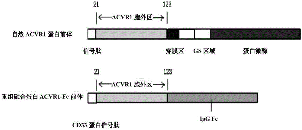
[0179] ACVR1-Fc expression gene consists of 3 fragments (such as image 3 shown), from the 5' end to the 3' end as follows:

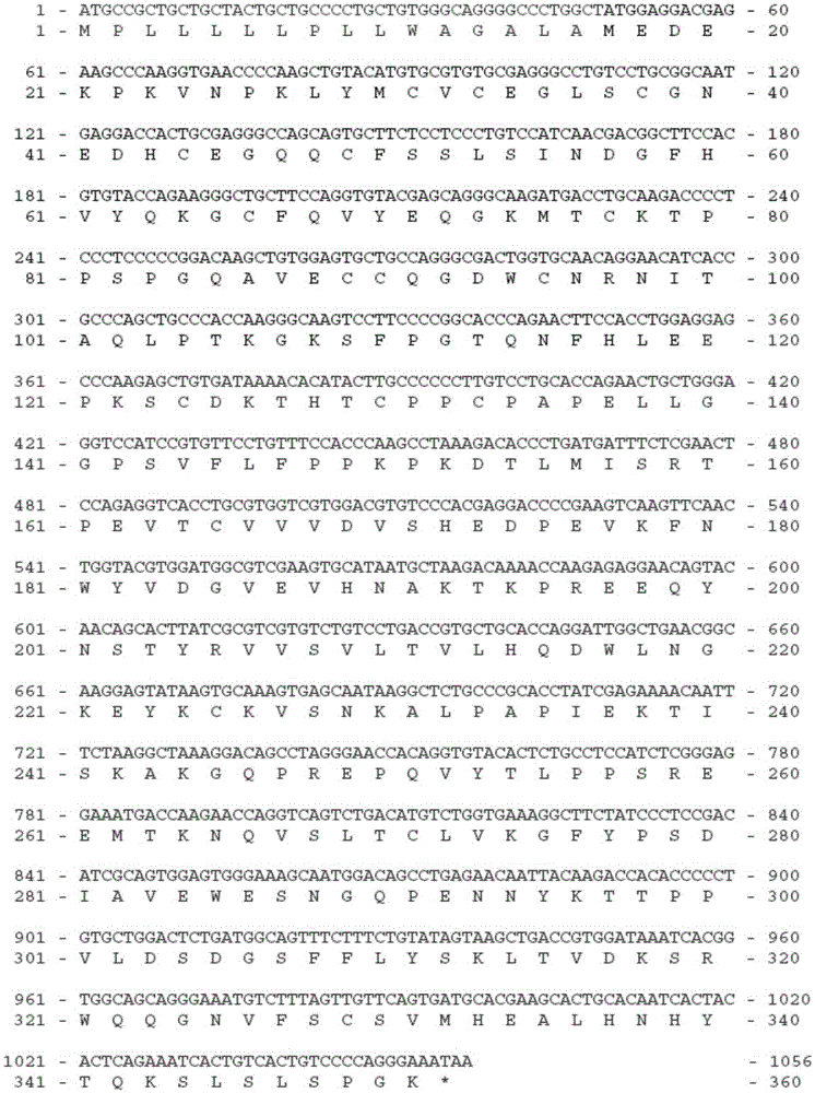
[0180] Fragment 1: the signal peptide sequence of the protein CD33 located at the 5' end (its coding sequence is shown in SEQ ID NO: 1, and its amino acid sequence is shown in SEQ ID NO: 2);

[0181] Fragment 2: the expression gene of amino acids 21-123 of the extracellular segment of ACVR1 located in the middle (its coding sequence is shown in SEQ ID NO: 3, and its amino acid sequence is shown in SEQ ID NO: 4);

[0182] Fragment 3: the coding sequence of the human IgGγ1 amino acid sequence at the 3' end (the coding sequence is shown in SEQ ID NO: 5, and the amino acid sequence is shown in SEQ ID NO: 6), which encodes amino acids from positions 216 to 447 of human IgGγ1 Residues, including the hinge region and the second and third CH regions (ie hinge region+CH2+CH3).

[0183] Th...

Embodiment 2

[0207] Example 2. Establishment of fusion protein expression cell lines

[0208] The host cell CHODG44 was derived from (purchased from Invitrogen Corporation, USA, Cat. No. 12609-012), and the method of cell culture and passage was referred to the CHODG44 manual of the company. Non-transfected cells were cultured in suspension in CDDG44 medium (Invitrogen) containing 8 mM L-glutamine and 5 μg / ml recombinant human insulin.

[0209] The CHODG44 cell line with stable and high-efficiency protein expression was established by stable transfection method. The cloned CHODG44 cells were cultured in suspension in serum-free, animal protein-free medium.

[0210] The method and steps of constructing a stable expression cell line of the fusion protein are briefly described as follows: use TianGen’s plasmid extraction kit to prepare the fusion protein expression vector plasmid, and digest 100 μg of the plasmid with the restriction endonuclease PuvI to make the plasmid linear. DG44 cell...

Embodiment 3

[0212] Example 3. Protein A affinity chromatography and HPLC-SEC analysis of fusion proteins

[0213] The ACVR1-Fc fusion protein was purified from the culture supernatant of stably expressing cells by protein A affinity chromatography. The purification method refers to the standard protein A (POROS, MabcaptureA) affinity chromatography method, and the purified protein is analyzed by reducing and non-reducing SDS-PAGE electrophoresis, and HPLC-SEC (high pressure liquid phase-molecular sieve) analysis.


PUM

PropertyMeasurementUnit
molecular weightaaaaaaaaaa

Abstract

The invention relates to a ACVR1-Fc fusion protein, and a preparation method and application thereof. Specifically, the invention relates to the fusion protein ACVR1-Fc containing an ACVR1 element and an Fc element, a DNA sequence coding the fusion protein, a vector containing the coding sequence, a host cell containing the vector, a preparation method for the fusion protein, and application of the above-mentioned substances in prevention and / or treatment of diseases or symptoms (such as diseases, cancers and the like related to pleonosteosis) related to ACVR1 abnormity (e.g., ACVR1 mutation and / or overactivity). The fusion protein provided by the invention has stable expression, high yield and high activity (e.g., effective inhibition of ossification and cartilage differentiation) and is simple to purify, so the fusion protein can be effectively used for prevention and treatment of diseases or symptoms related to ACVR1 abnormity.

Description

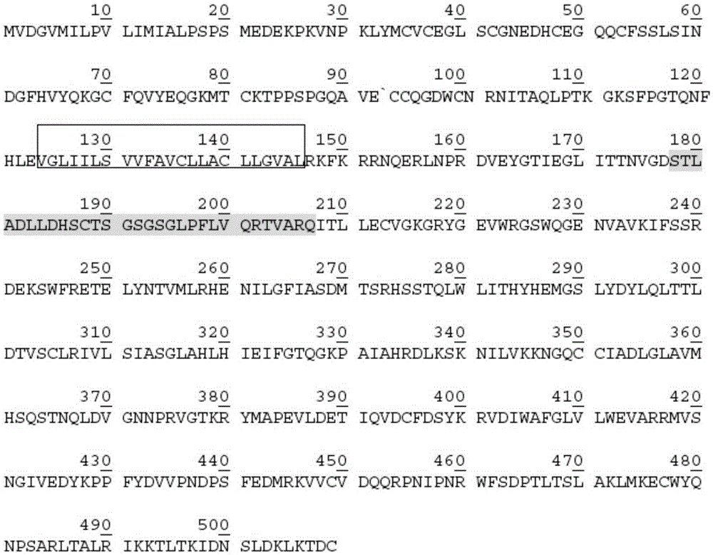
technical field [0001] The invention belongs to the fields of biotechnology and medicine. Specifically, the present invention relates to activating protein A receptor 1-Fc (activinAreceptor, typeI-Fc, ACVR1-Fc) fusion protein, its preparation method and its role in the prevention and / or treatment of ACVR1 abnormalities (such as ACVR1 mutations and / or Hyperactivation)-related diseases or symptoms (eg, hyperossification-related diseases, diffuse intrinsic pontine glioma, ovarian cancer, etc.). Background technique [0002] ACVR1, one of the subtypes of bone morphogenetic protein (BMP) type I receptor, Activin receptor type IA (ActRIA), is also known as Activin receptor-like kinase 2 (Activin receptor-likekinase2, ALK2). [0003] ACVR1 belongs to transforming growth factor β (TGF-β) superfamily receptor type I, and the whole receptor protein is composed of extracellular segment, transmembrane segment and intracellular segment. The C-terminus of the intracellular segment is a ...

Claims


Application Information

Patent Timeline
Patent Type & Authority Applications(China)
IPC IPC(8): C07K19/00C12N15/62C12N15/63A61K48/00A61K38/17A61P35/00
CPCA61K38/00A61P19/08A61P35/00C07K14/71C07K19/00C07K2319/02C07K2319/30C12Y207/1103
Inventor 陈羿蔡则玲张克勤
Owner SHANGHAI KANDA BIOTECHNOLOGY CO LTD
Learn More