Method for determining radiosensitivity

A sensitive, compound technology, applied in the direction of radiotherapy, X-ray/γ-ray/particle irradiation therapy, treatment, etc., can solve the problem of not having both positive and negative predictive value, lack of technical feasibility, lack of sensitivity and/or specificity sexual issues

Inactive Publication Date: 2016-01-20
蒙彼利埃大学医疗中心 +2
View PDF4 Cites 4 Cited by
  • Summary
  • Abstract
  • Description
  • Claims
  • Application Information

AI Technical Summary

Problems solved by technology

Finally, Oh et al suggested that the original and identification of α-2 macroglobulin in silico assays might be associated with an increased risk of radiation-induced lung inflammation in lung cancer patients, but did not confirm this in an independent cohort. Results (Oh et al., 2011)
[0008] None of the existing predictive tests for radiosensitivity are available for clinical routine due to two major drawbacks: i) they lack sensitivity and / or specificity, do not have both good enough positive and negative predictive values, and ii) Its lack of technical feasibility and long delays require trained practitioners, its high cost and invasive collection of biological samples

Method used


Image

  • Method for determining radiosensitivity
  • Method for determining radiosensitivity
  • Method for determining radiosensitivity

Examples


Embodiment 1

[0148] Example 1: Lymphocyte apoptosis analysis

[0149] The inventors previously developed a rapid and reproducible assay called RILA (Radiation-Induced Lymphocyte Apoptosis), which measures apoptosis of CD4 and CD8 T-lymphocytes after radiation (0.5-8 Gy) by flow cytometry. This measurement is based on the reduction of nuclear DNA fluorescence resulting from specific chromatin changes that accompany apoptosis. In 150 patients, RILA was used as the primary stratification factor in a phase II randomized study of early breast cancer following conventional surgery compared with concurrent or sequential postoperative radiation therapy with letrozole. point) is breast fibrosis (Azria et al., 2010). Patients with RILA >16% were not found to exhibit radiation-induced late effects, indicating a high negative predictive value of this test. All patients with grade 2 or worse subcutaneous fibrosis had <16% RILA, confirming the predictive value of the test. However, among patients wit...

Embodiment 2

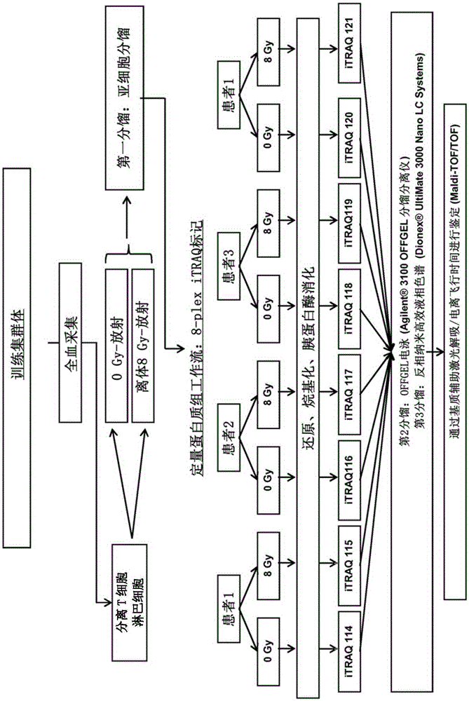
[0150] Example 2: Identification of predictive markers of late-induced cytotoxicity

[0151] The protocol used to identify predictive markers is shown in figure 1 middle. From the aforementioned four patients, a total of 40 ml of blood was collected in heparinized tubes. Use the rosette (rosette) according to the manufacturer's instructions ( StemCell Technology) performed negative selection followed by isolation of T-lymphocytes from whole blood by Ficoll gradients (GE Healthcare). Lymphocytes were then cultured in RPMI medium with 10% FCS at 37°C and 5% CO2 for 24 hours. Subsequently, half of the lymphocytes were irradiated with 8Gy in vitro. Then, irradiated and non-irradiated lymphocytes were cultured again for 48 hours at 37°C and 5% CO2. After this incubation time, lymphocytes from each patient were then submitted to subcellular fractionation (fraction) ( The Subcellular Proteome Extraction Kit (cat. 539790, Merckmillipore) allows the separation of cytoplasmic, m...

Embodiment 3

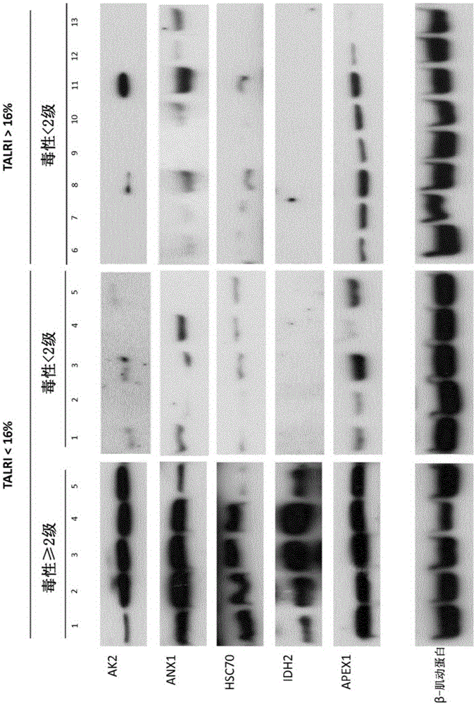
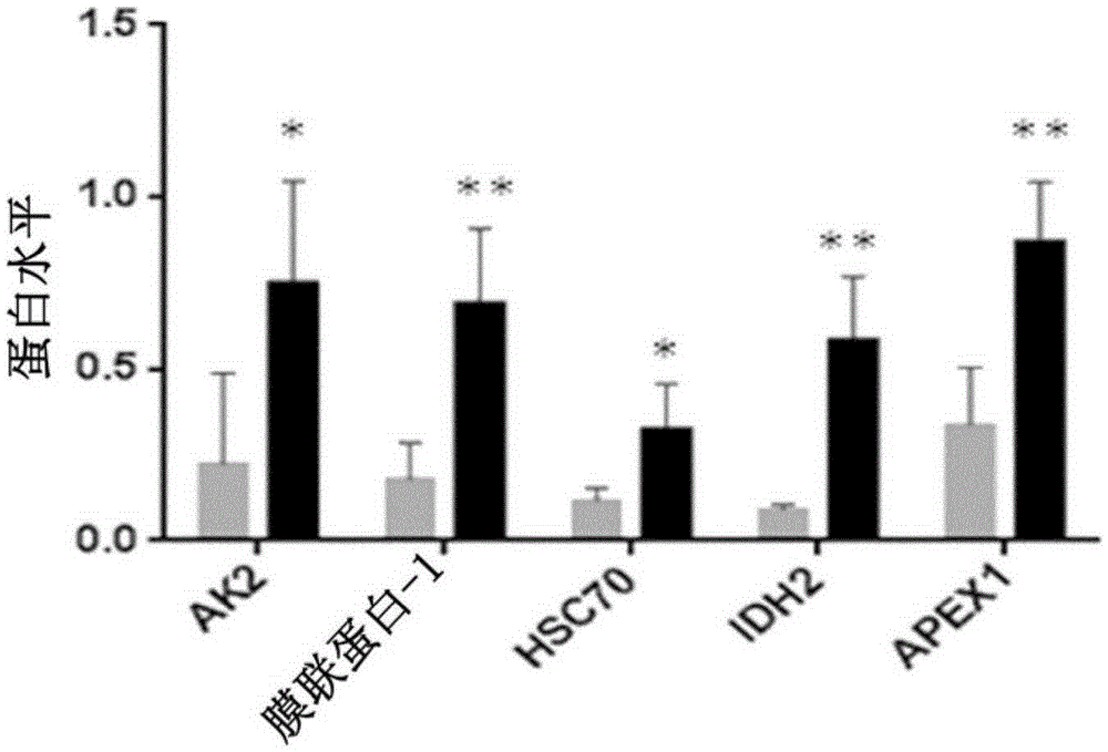
[0156] Example 3: Confirmation of differential expression of biomarkers in a larger number of patients following radiation therapy

[0157] These five proteins were validated by western blot analysis in an additional population of 18 patients, of which 5 patients had developed > grade 2 breast fibrosis and 13 patients had only mild or no toxicity. All these 10 patients exhibited low RILA values. Blood samples were collected and processed until post-irradiation incubation as described in previous examples. Then, lymphocytes were lysed in RIPA buffer. Then, the protein was quantified again, and each 10 μg was placed in a 12% polyacrylamide gel for Western blotting. After migration, transfer to PVDF membrane at 300mA for 1 hour at 4°C, then saturate the membrane for 2 hours in PBS-Tween 0.05%-milk 5%, the antibody against the protein of interest in the same saturation buffer in Incubate overnight at 4°C with agitation. After five consecutive 5 min washes in PBS-Tween 0.05% bu...


PUM


Abstract

The present invention relates to a method for the in vitro determination of the radiosensitivity of a subject. More particularly, the invention relates to a method comprising a step of inducing an exogenous stress on a biological sample from a subject, followed by the comparison of the presence or level of at least one compound chosen in a group of defined compounds, in said biological sample and in a reference sample. The present invention also relates to the use of said at least one compound as predictive biomarker of the radiosensitivity of a subject. The invention also relates to a kit for the detection of the presence or level of at least one of said compounds, usable in a method according to the invention.

Description

technical field [0001] The present invention relates to methods for determining radiosensitivity of a subject in vitro. More specifically, the present invention relates to a method comprising inducing exogenous stress in a biological sample from a subject and comparing the level of at least one identified compound between said biological sample and a reference sample. The present invention also relates to the use of said at least one compound as a predictive biomarker of radiosensitivity in a subject. The present invention also relates to a kit for detecting the level of at least one of said identified compounds that can be used in the method according to the invention. Background technique [0002] The success of radiation therapy is largely dependent on the total administered dose. Individuals vary widely in the susceptibility of tissues to injury from ionizing radiation. Worldwide, approximately 4 million people are treated with radiation therapy each year. Current es...

Claims


Application Information

Patent Timeline
Patent Type & Authority Applications(China)
IPC IPC(8): A61N5/10G01N33/574
CPCA61N5/103G01N33/6893G01N2800/40G01N2800/50G01N2800/60G01N2800/7004
Inventor D·阿兹里亚J·拉孔巴J·索拉索尔A·曼格
Owner 蒙彼利埃大学医疗中心
Why Eureka
Try Eureka