T-shaped positioning device adopting 3D printing in intrathoracic endoscopy and manufacturing method thereof

A positioning device and 3D printing technology, applied in surgery, medical science, computer-aided planning/modeling, etc., can solve problems such as dyspnea, rib/scapula affecting positioning, difficult nodules, etc., to reduce workload and reduce exposure The effect of measuring and avoiding pneumothorax

Active Publication Date: 2016-07-27
SHANGHAI PULMONARY HOSPITAL
View PDF7 Cites 9 Cited by
  • Summary
  • Abstract
  • Description
  • Claims
  • Application Information

AI Technical Summary

Problems solved by technology

[0004] However, the existing technology may have the following problems: the mass cannot be touched by manual palpation: the diameter of the lung ground glass nodule is too small, the depth from the subpleural depth of the visceral layer is too deep, or the texture is soft and difficult to distinguish from the surrounding normal lung tissue, which greatly increases the difficulty of manipulation. The difficulty of palpation makes it difficult to locate the nodules accurately, which eventually leads to the inability to completely resect the pulmonary nodules; ribs / scapulas affect positioning: due to anatomical reasons, pulmonary ground-glass nodules (GGO) in the lung tissue covered by ribs / scapulas cannot pass through the body Positioning by puncture; positioning device falling: For small nodules that are relatively close to the visceral pleura and shallow, the hook-wire positioning needle and micro-coil micro-coil are used for lung puncture positioning because the puncture is too shallow. Incomplete penetration into the lung parenchyma may easily cause the positioning needle or coil to fall off and fall into the thoracic cavity; pneumothorax: the hook-wire positioning needle or micro-coil micro-coil is used for lung puncture, and sometimes repeated punctures are required for accurate positioning. It is easy to cause lung rupture and cause pneumothorax, and the patient has symptoms such as chest tightness, shortness of breath, and dyspnea, or the blood vessel is accidentally injured by the positioning needle, causing bleeding, or even hemothorax, and the patient has hemorrhagic shock, such as pulse speed and blood pressure drop. Effects of radiation radiation: preoperative Puncture positioning guided by CT via hook-wire positioning needle or micro-coil microcoil requires multiple CTs in a short period of time to determine the positioning point, which increases the radiation dose received by patients and doctors

Method used


Image

  • T-shaped positioning device adopting 3D printing in intrathoracic endoscopy and manufacturing method thereof
  • T-shaped positioning device adopting 3D printing in intrathoracic endoscopy and manufacturing method thereof
  • T-shaped positioning device adopting 3D printing in intrathoracic endoscopy and manufacturing method thereof

Examples


preparation example Construction

[0045] The present invention also provides a method for preparing a T-shaped positioning device based on 3D printing in endoscopic thoracic surgery of pulmonary ground-glass nodules, comprising the following steps:

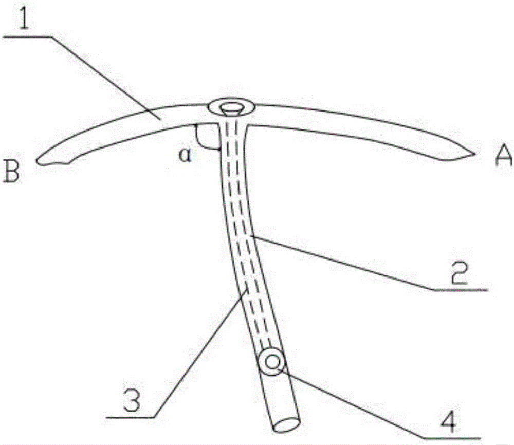
[0046] (1) Based on the pre-processed images of localization slices with lung masses, use modeling software to reconstruct the model of the patient's chest and lungs with pulmonary ground-glass nodules, and mark the lung ground-glass on the chest and lung models the location of the nodule;

[0047] (2) Using the modeling software again to construct a fitted "T"-shaped structural model on the chest cavity and lung models according to the preset minimally invasive incision, which includes a horizontal section and a vertical section;

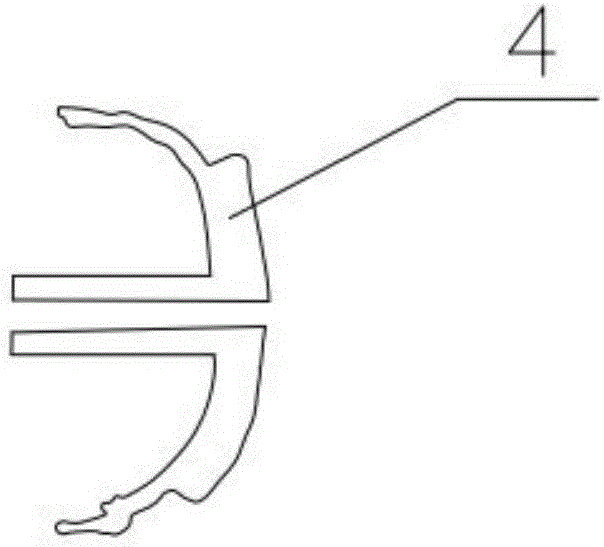
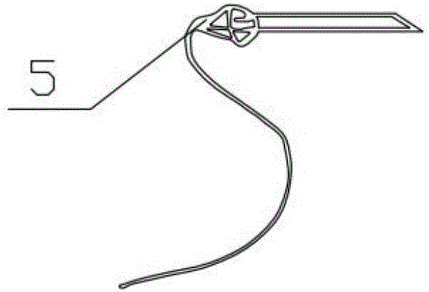
[0048] (3) Import the "T"-shaped structural model into the printer and perform 3D printing, and install a lesion marking device connected to the hollow pipe at the corresponding position on the printed vertical section to obtain a "T" ...


PUM


Abstract

The invention discloses a T-shaped positioning device adopting 3D printing in an intrathoracic endoscopy and a manufacturing method thereof. The T-shaped positioning device is of a T-shaped structure adopting the 3D printing and comprising a vertical section and a horizontal section, wherein the upper end of the vertical section and the middle of the horizontal section are intersected at a certain angle, a hollow tube communicated with the surface of the horizontal section is arranged in the vertical section, the lower end of the vertical section is closed, and a nidus marking device connected with the hollow tube is arranged on the vertical section. When the T-shaped positioning device is used, the positioning device is arranged and fixed in the thorax of a patient in a conformal mode and marks the ground-glass node position of the lung through the nidus marking device. The T-shaped positioning device avoids the problem that a node and a hook-wire cannot be touched by adopting a method of touching by hand or a micro-coil is unhooked or falls into the thoracic cavity, the CV exposure to the patient and the working amount of a medical worker are decreased, and meanwhile the influence on the ribs or shoulder blades due to dissection is eliminated.

Description

technical field [0001] The invention relates to a method and a device for determining pulmonary ground-glass nodules in the thoracic cavity, in particular to a 3D printed T-shaped positioning device for thoracic endoscopic surgery and a preparation method thereof. Background technique [0002] Pulmonary ground-glass nodules are mildly increased densities on CT images, but the bronchovascular bundles within them can still be displayed. This sign is often the manifestation of early lung disease. Timely detection and diagnosis are of great significance for correct clinical treatment and prognosis. It is an indication for laparoscopic surgery. However, because the lesion is small and the chest cavity is an internal cavity, Pulmonary ground-glass nodules are difficult to pinpoint precisely because lung tissue and the thorax move with breathing. [0003] At present, there are three main methods for determining pulmonary ground-glass nodules (GGO): 1. Intraoperative exploration an...

Claims


Application Information

Patent Timeline
Patent Type & Authority Applications(China)
IPC IPC(8): A61B90/00A61B17/00A61B34/10
CPCA61B17/00234A61B2017/00526
Inventor 王轶灵张旭峰李牧李泽遥王龙范子文顾亚伟陈昶
Owner SHANGHAI PULMONARY HOSPITAL
Why Eureka
Try Eureka